We decided on placing an implant in the upper left 4 position of a male English patient after taking a panorama x-ray. His tooth had been extracted 3 months previously, the space was relatively fresh. The implant: Pitt Easy V-TPS. 4*10. We gave him a medicine pack (Dalacin antibiotics and Cataflam painkillers), and ice-gel pack.
One of our Irish patients had previously lost two front teeth which we replaced with implants 3 months ago. Today we are already fitting the abutments. In order to protect the restoration we have recommended producing a bite raising nightguard which the patient has agreed to. Therefore we also took impressions for the bite raising nightguard. Because this can be produced in a short space of time, it fits into the patient’s stay in Hungary.
After missing teeth for a long time, our Danish patient will receive new teeth in positions upper right 4 and 6 with the help of implants. Following the sinus lift we placed the implants, and gave the patient antibiotics and painkillers (Dalacin C and Cataflam). The patient did not require a temporary restoration. In the next stage – in 4 months time – the patient will receive their teeth.
A 63 year old female patient wants implants in positions upper right 3, 5 and 6, because teeth have been missing in these positions for a long time. The lady’s sinus cavity was dilated thus the bone layer needed to support the implants is too thin.
An American female patient presented with multiple missing teeth and an unremarkable resorbtion in the jaw ridge. In order to be able to place implants in positions 23,24,25,26 it was necessary to carry out a minimal sinus lift. The sinus lift is a surgical treatment which we usually carry out with local anaesthetic. In the case of this patient we used BioOss, a bone replacement material of animal origin. Because the sinus lift was not significant we carried out the bone replacement and placing of implants in only one session. We also used BioGide membrane to help the tissue to heal quicker. The collagen membrane, also of animal origin, helps the wound to heal and has a positive effect on bone regeneration as well. We will remove the patient’s stitches next week. Then a 6 month osseointegration period is necessary before we will finally be able to place the new porcelain teeth. Unusually, the lady did not want a temporary denture/crowns for the interim period.
A returning middle-aged Hungarian gentleman complained of sensitive gums which bled easily when pressure was applied in the area of tooth 47 where a dental implant was inserted many years ago. During the examination we diagnosed inflammation and periimplantitis around the implant.
The inflammation around the implant had penetrated to deeper levels and had caused the bone tissue to disappear. The cause of an infection of this nature is usually bacterial. In the pockets which had developed between the dental implant and mucosa, with a lack of regular and thorough mouth and tooth care the bacteria can easily colonize and cleaning off the tartar can be difficult. If the infection also reaches the bone tissue the implants can loosen and if left untreated the patient may lose them. Our patient’s inflammation around the implant was indeed at an advanced stage. The tissue decay had loosened the implant as well therefore we removed it. After proper cleansing of the wound we carried out a new implantation. The patient will get a new crown after the healing period.
A 26 year old patient came to us because 2 of his incisors had broken in a childhood accident and were built up with composite fillings. According to him this solution is aesthetically not satisfactory and he would rather have crowns. We took a panorama x-ray and provided a treatment plan. The patient accepted the plan. By grinding down the teeth, we prepared the 2 incisors for zirkonium crowns and took a precision impression.
We fitted zirkonium crowns to a young Norwegian patient’s upper front teeth (from 13 to 23). Aesthetics were a very important factor for the young man, therefore he chose the more expensive but more aesthetic zirkonium, while we resolved his other missing teeth (lower and upper, 4 to 7) with porcelain crowns fused to metal. The restoration was functionally and aesthetically successful, and the patient satisfied.
A middle aged patient returned to our clinic because of damage to ceramic units of a previous placed bridge. The damage occurred a night therefore, in order to protect the upper and lower round bridge, we recommended the patient wear a bite-raising nightguard. Because this is not an extreme case of denture grinding, a thin guard is sufficient. We took an alginat impression for the nightguard. The production of the guard takes 1-2 days.
A 52 year old Norwegian presented for the metal frame tryout. An upper porcelain fused to metal restoration will be produced from upper right 7 to upper left 7. We determined the tooth colour which was agreed with the patient. Because there are natural teeth missing over the whole length of the upper, it was possible to choose a tooth colour differing somewhat from the original colour. The lady’s lower jaw also lacked teeth therefore we will place two lateral bridges in the lower region at some point.
A middle aged man presented with 1 tooth missing from the upper left side. For the first stage of treatment we finished the preparation (grinding), took the impression and placed the temporary crowns. At the present stage we tried the metal frame for the porcelain bridge fused to metal, and we produced a model of the new bite with Futar-D. The test was successful and and the height perfect.
One of our American patients presented a few days previously with dental restorations in a bad condition. His/her expectationwas that we would produce an upper and lower round bridge on his/her existing implants. At the first treatment we prepared the teeth in the lower jaw, grinded them down, exposed the implant sites and took a precision impression. Today we prepared the left 3 in the upper jaw. We exposed the implants existing int hat region and placed impression caps into them. We then took a precision impression. Following the production of the round bridges at the next treatment the round bridges were fitted to the upper and lower implants. After permanently fitting the bridges, the patient could smile again!
A 23 year old Irish patient presented following many previous dental treatments. We found 3 well placed implants in the patient’s upper jaw, but the abutments and many of the young lady’s own teeth were in an eroded and abraded condition. We fitted zirconium and porcelain fused to gold crowns and bridges, and glass fibre post cores, to the implants and the patient’s own teeth. Here we were able to save 2 roots which were still in a good condition. The patient’s teeth show that during previous treatment acute complaints were indeed carefully attended to but no attempt was made to solve the probable causes of the problems: nocturnal grinding (bruxism). On our recommendation the young lady also received a nightguard (bite raising splint for night time use).
An upper porcelain fused to metal round bridge with post cores was produced swiftly within 1 week in the dental technical laboratory for a 60 year old English female patient. For the fitting and cementations we used FujiPlus cement. The height of the bite was fine. We took an alginate impression for the patients bite raising nightguard. The patient can collect the nightguard in 2 days’ time. We provide the patient with further oral hygiene advice. She needed this on the basis of the condition of the oral cavity. The patient was very satisfied with the treatment and had the opportunity to see the sights of Budapest whilst waiting for her new teeth.
A 32 year old Norwegian lady came to us following her second pregnancy as her dentition had deteriorated significantly following the births. We prepared all of her upper teeth, took impressions of the tooth stumps and produced a temporary bridge.
The patient wanted full porcelain crowns, where the crown frame is also made from zirconium-oxide. In this instance we produce the frame using 3D computer scanning techniques which enables better precision and aesthetic results in the finished product. The laboratory procedure is therefore more time consuming and the materialas well as the method make the process more expensive but the look of the crowns is very important to this Norwegian patient.
We root canal treated the upper left 2 tooth of a 40 year old French male patient. We determined the length of the root canal with electronic measuring equipment. The root canal required mechanical … . Following the disinfection and drying of the root canal, we used Sealapex for the root filling and Fuji IX cement for the cover filling.
One of our Norwegian male patients complainedof a recurring sometimes splitting toothache. During the diagnosis we established that tooth 32 was significantly decayed and a root canal treatment would be necessary. During the treatment the root canal of tooth 32 was opened up and cleaned using WDW Gold. During the rinsing of the root canal we used Solumium then placed a medical (calcipulp) temporary filling. As expected the patient’s pains subsided and disappeared gradually after a few days. If everything goes well we will change the temporary root filling for a permanent one in 8 days’ time.
A Hungarian patient presented with swollen gums. 2 months ago the patient received an implant in the upper right 4 position. The swelling was between the upper right 4 and 3. We checked the implant by taking an x-ray and found everything to be in order. In order to disinfect the swelling we cleaned the swollen area with Betadin. It is likely that the patient scraped the gum with some sort of hard food because we did not detect any pathological mutation.
One of our Hungarian patients was treated 3 weeks ago in Germany. The lower left 8 (wisdom) tooth was removed because, according to the patient’s account, there was a cyst around it. The patient presented at our clinic with a swollen face and complaining of a sharp throbbing pain. We rinsed the area with Hyperol Betadin and removed bone splinters from the wound. We finally treated the patient with antibiotics. As all bone splinters have been successfully removed afterwards, the pain will subside within a short period of time and the swelling will go down.
A 31 year old man presented with a headache which had been getting worse for days. Additionally, discolouration was visible with the naked eye on the lower first molars. The patient’s diagnosis was correct, we were faced with cavitied teeth. In the lower right and left number 4 teeth we prepared photopolymer fillings which have the advantage of bonding immediately through light. It is not necessary to wait even 1 to 2 minutes, and it possesses outstanding aesthetic properties.
A young male Norwegian patient had been sleeping badly for a long time, waking up tired, with a headache and stiff neck. Additionally his gum was regularly swollen and his jaw muscles hurt which is indicative of his sleeping the whole night with clenched teeth. This is also supported by the fact that we found signs of wearing in some places on his teeth. We produced for the patient a nightguard which will prevent the constant clenching of his teeth and will protect his teeth from abrasion.
A 29 year old patient complained of an annoying but not unbearable tooth pain which had been present for several days. We found deep decay in the upper right 3 tooth where we used Dycelal disinfectant in the indirect capping of the pulp, which we covered with Fuji II.LC glass ionomer cement.
The ortho-pantograph x-ray of our 50 year old Italian femaile patient showed that her upper right 6 and upper left 6 (or rather their remains) needed extracting and replacing. The extracted teeth were replaced with bridgework according to the plan. In the first stage of treatment we prepared the right and left upper 5 and 7 teeth, and removed the right and left upper roots (radixes). We took impressions for the dental laboratory and produced temporary crowns. The dental technical laboratory will produde the final crowns in under 1 week and the Italian lady’s next visit will be due in only 2 weeks’ time. This way she was able to purchase an economy flight ticket.
A young Swiss man complained of a sharp throbbing pain in his lower left teeth. The problem was caused by the severely inflamed 37. We trepanated and extirpated this number 7, which means we opened the pulp chamber and removed the inflamed nerves. We carried out the widening and cleaning with WDW Gold. We placed a Sealapex root filling and then a Fuji IX tooth filling.
A 60 year old English gentleman presented with many problems. Firstly we surgically removed teeth 12,44,45 and 48. We produced the treatment plan for the replacement of the teeth but he did not want to begin the restoration treatment on this visit.
Secondly in the right upper and lower quadrants it was necessary to remove tartar. Following the ultrasound treatment and cleaning we carried out a curettage in the deeper parts of the gum pockets. This is necessary because the deeper parts of the gum pockets are not reachable with ultrasound scaling. With the help of the appropriate instruments these areas can be cleaned for which local anaesthetic is usually required. On finishing treatment we provided our patient with oral hygiene advice for which the patient was especially grateful.
Kreativ Dental Clinic © 2025
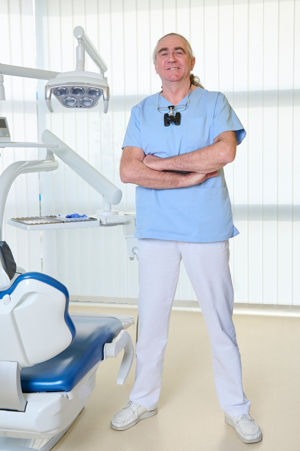
Cristian graduated in 1988 from the Neumarkt Dental School. In 1992 he became a specialist in General Dentistry, and since 1994 has pursued his particular interest in Implantology and Cosmetic Dentistry. Cristian has also achieved the Cambridge First Certificate in English.
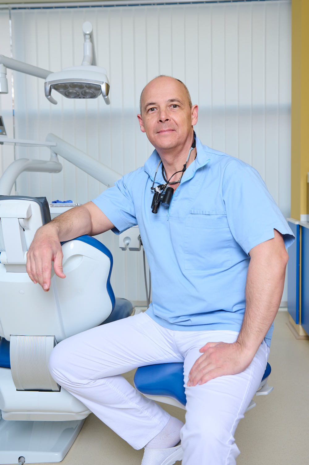
András first qualified in 1992 as a General Dentist from Semmelweis Medical University, Budapest. After becoming a specialist in General Dentistry in 1995, he worked as a private dentist and practiced as a dental surgeon at Rókus Hospital, Budapest. In 1995 he became a specialist in General Dentistry. His specialist field is Crown and Bridgework.
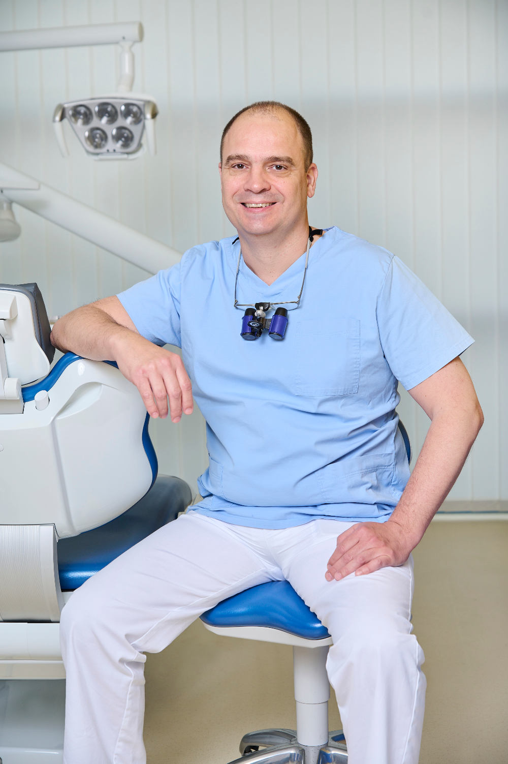
Balázs qualified in 1999 from Semmelweis Medical University as a General Dentist. He then worked for 2 years as a Clinical Dentist. After 6 years experience treating German and Austrian patients in Hungary, he joined Kreativ Dental in 2007. Balázs speaks both English and German.
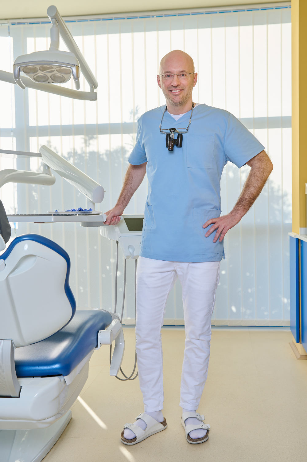
Iván qualified in 2000 from Semmelweis Medical University, Budapest. In 2002 he joined Kreativ Dental after gaining 2 years experience in private practice. His special interests are Endodontics and Cosmetic Dentistry
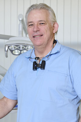
Péter qualified in 1994 from the University of Dentistry, Budapest. He was awarded a Fellowship in Dental Surgery for two years at the University of Dentistry in Vienna. Following several hospital positions in restorative dentistry and oral-maxillo facial surgery where he gained valuable surgical training, he joined our dental team in 1998. His special interest is Cosmetic Dentistry.
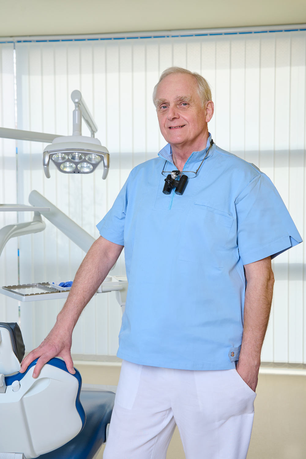
Louis first qualified in 1989 as a General Doctor from Semmelweis Medical University, Budapest. In 1994 he qualified as a General Dentist, qualifying in Maxillo-Facial Surgery in 1999. Since then he has been working exclusively as a Dental Surgeon and his specialist field is Implantology.
Medical Registration Number: 48939
Numéro d’inscription médicale : 48939
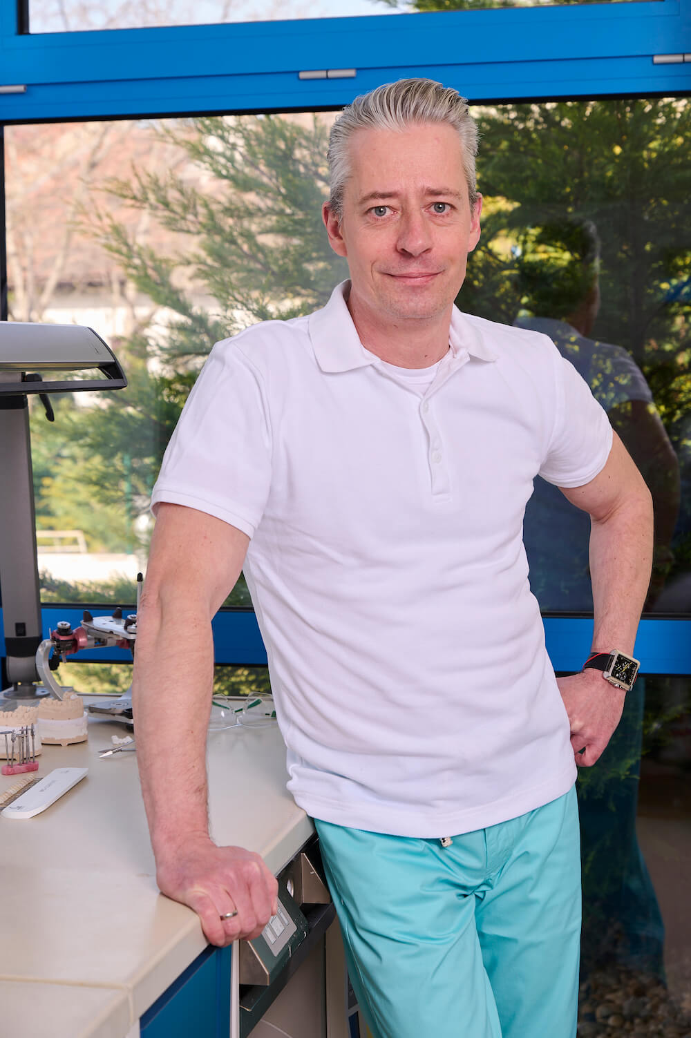
Keve Horváth joined our team of dental technicians. He works in the Ceramic Department as a master ceramist. After he graduated in 1994, he furthered his knowledge from several symposiums in Hungary, Germany and Belgium (for example Noritake and Ivoclar). In 2006 he held a course „Esthetic Front” with a real patient presence, which is a special event in Hungary and also in Europe.
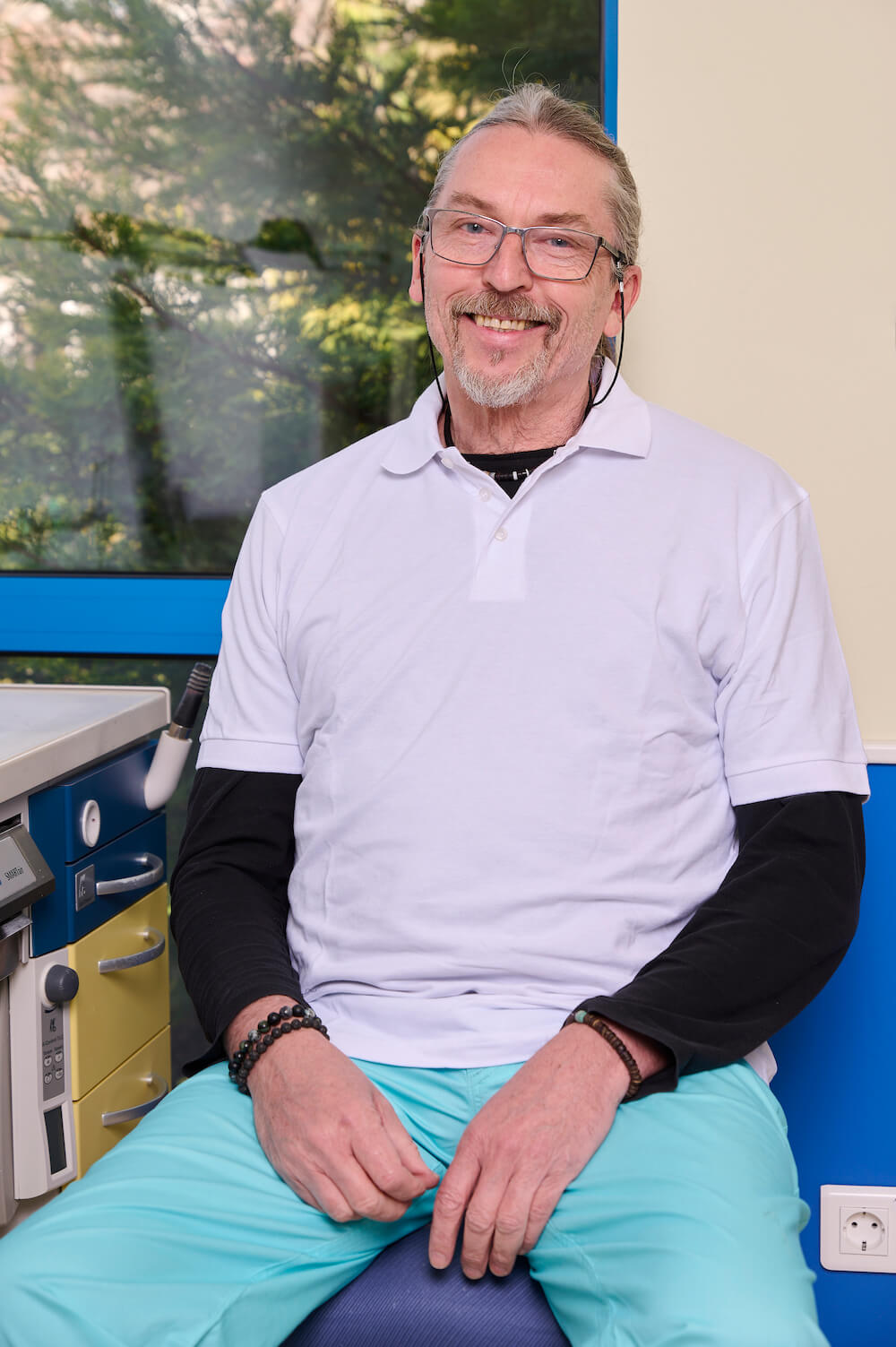
János is Leader of the Ceramic Department where all Porcelain Fused to Metal Crowns, Full Porcelain Crowns, Veneers and Inlays are skillfully crafted. Furthermore, he is one of the few exclusive demonstrators in Europe of the German company Vita which produces the best quality dental porcelain in the world.
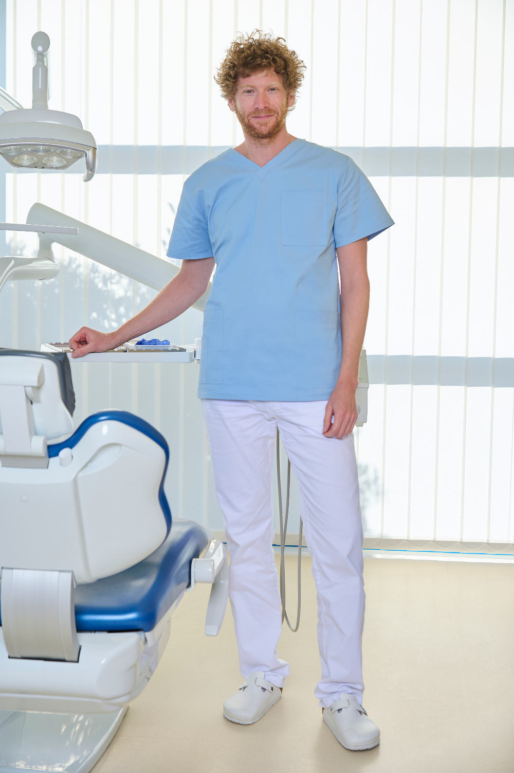
Ádám graduated in 2011 from the Semmelweis University, Faculty of Dentistry. Since then he worked at the Department of Conservative Dentistry, untill he finished a 3 year postgraduate training in conservative dentisty and prosthodontics. He also participated in the education of students both in Hungarian and English language. Ádám joined the Kreativ Dental team in 2014. His special interest is endodontics.
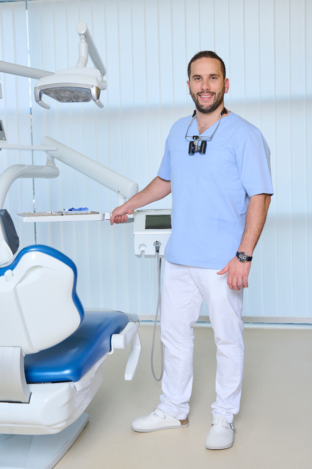
Bernard graduated in 2012 from the Semmelweis University, Faculty of Dentistry. Since then he has worked at the Department of Maxillofacial Surgery at St. John’s Hospital Budapest for 3 years and then obtained his specialization in dentoalveolar surgery in 2015. Meanwhile, he worked in private dentistry as a general dentist.
His special field is oral surgery, implantology and prosthodontics.
He joined the Kreativ Dental team in 2015.
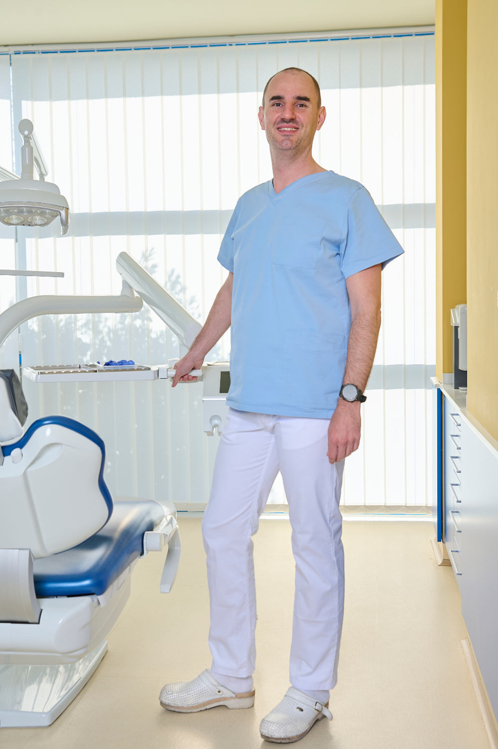
Bálint qualified at Semmelweis University, Faculty of Dentistry in 2007. Since then he has been working at the Department of Periodontology, Semmelweis University, and participates in national and international conferences (Europerio, IADR). He finished his 3 year postgraduate periodontal training in 2010. Bálint joined Kreativ Dental in 2013, his specialist field is periodontal surgery.
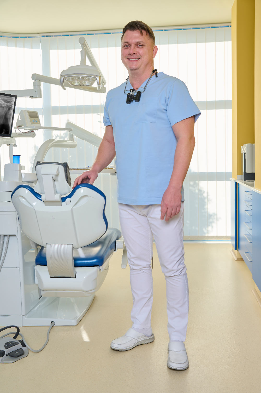
Ádám graduated in 1998 from the University of Szeged. In 2000 he became a Dental Surgeon /Specialist in General Dentistry.
After working as a Dental Surgeon at BKKMi County Hospital, Maxillo-Facial Department, Kecskemét, Hungary between 2000 and 2006, he spent 7 years as private dentist and as General Dental Practitioner in the United Kingdom.
He joined the Kreativ Dental team in 2013.