Vi bestämde oss för att sätta in ett implantat i vänster överkäke position 4 på en manlig engelsk patient efter att ha tagit panorama röntgen. Hans tand hade dragits ut 3 månader tidigare, så det var ett relativt färskt område. Implantatet: Pitt Easy V-TPS. 4 * 10. Vi gav honom en läkemedelsförpackning; (Dalacin antibiotika och Cataflam smärtstillande medel) och is-gel pack.
En av våra irländska patienter hade tappat två framtänder som vi ersatte med implantat för 3 månader sedan. Idag ska vi montera distanser. För att bevara och skydda den färdiga restaureringen har vi rekommenderat en bettskena att användas på natten. Därför tog vi avtryck för bettskena, och eftersom en sådan kan tillverkas på kort tid, hinner vi få den färdig medan patienten är i Ungern..
Efter att ha saknat tänder under en längre tid, ska vår danska patient få nya tänder i position 4 och 6 i överkäken med hjälp av implantat. Efter sinus lift (käkbensförstärkning) satte vi in implantat, och gav patienten antibiotika och smärtstillande medel (Dalacin C och Cataflam). Patienten tyckte inte att han behövde temporära tänder. I nästa steg – ca 4 månader senare – ska patienten få sina tänder.
En 63 årig patient vill ha implantat i positon 3, 5 och 6 i höger överkäke där hon sedan länge saknat tänder. Hennes käkben har blivit för tunt, och måste förstärkas genom sinus lift för att få stabilt fäste för implantat.
An American female patient presented with multiple missing teeth and an unremarkable resorbtion in the jaw ridge. In order to be able to place implants in positions 23,24,25,26 it was necessary to carry out a minimal sinus lift. The sinus lift is a surgical treatment which we usually carry out with local anaesthetic. In the case of this patient we used BioOss, a bone replacement material of animal origin. Because the sinus lift was not significant we carried out the bone replacement and placing of implants in only one session. We also used BioGide membrane to help the tissue to heal quicker. The collagen membrane, also of animal origin, helps the wound to heal and has a positive effect on bone regeneration as well. We will remove the patient’s stitches next week. Then a 6 month osseointegration period is necessary before we will finally be able to place the new porcelain teeth. Unusually, the lady did not want a temporary denture/crowns for the interim period.
A returning middle-aged Hungarian gentleman complained of sensitive gums which bled easily when pressure was applied in the area of tooth 47 where a dental implant was inserted many years ago. During the examination we diagnosed inflammation and periimplantitis around the implant.
The inflammation around the implant had penetrated to deeper levels and had caused the bone tissue to disappear. The cause of an infection of this nature is usually bacterial. In the pockets which had developed between the dental implant and mucosa, with a lack of regular and thorough mouth and tooth care the bacteria can easily colonize and cleaning off the tartar can be difficult. If the infection also reaches the bone tissue the implants can loosen and if left untreated the patient may lose them. Our patient’s inflammation around the implant was indeed at an advanced stage. The tissue decay had loosened the implant as well therefore we removed it. After proper cleansing of the wound we carried out a new implantation. The patient will get a new crown after the healing period.
En 26-årig patient kom till oss därför att 2 av hans framtänder hade slagits sönder vid en olycka i barndomen och hade lagats med kompositfyllning. Nu tyckte han inte att det såg bra ut utan ville hellre ha kronor. Vi tog panorama röntgen och gav en behandlingsplan, som patienten accepterade. Vi förberedde tänderna för zirkonium porslinskronor genom att slipa dem och tog därefter precisionsavtryck.
Vi monterade zirkonium kronor på en ung norsk patients 6 främsta tänder i överkäken. Estetik var en mycket viktig faktor för den unge mannen, därför han valde den dyrare men mer estetiska helporslinskronan. Övriga saknade tänder (nedre och övre, 4 till 7) ersatte vi med metallkeramiska kronor. Restaureringen var funktionell och estetiskt lyckad och patienten blev nöjd.
En medelålders patient återvände till vår klinik på grund av skadade porslinskronor på en bro, som vi satt in tidigare. Skadan inträffade under natten. För att skydda de båda bryggorna i över- och underkäke, rekommenderade vi patienten att ha bettskena nattetid. Eftersom detta inte var ett extremt fall av tandgnissling, var en tunn skena tillräcklig.Vi tog ett alginatavtryck. Tillverkningen av bettskena tar 1-2 dagar.
Metallram ska provas ut på en 52-årig norska. En metallkeramisk brygga från tand 7 i vänster överkäke till tand 7 i höger överkäke ska tillverkas. Tillsammans med patienten bestämmer vi färg. Eftersom alla tänder saknas i överkäken kan färgen få bli lite annorlunda jämfört med den ursprungliga. Kvinnan saknar även tänder i underkäken där två bryggor ska att sättas in.
En medelålders man saknade en tand i vänster överkäke. Behandlingen inleddes med att slipa intilliggande tänder, ta avtryck och sätta in temporära kronor. Här utprovar vi metallramen till den metallkeramiska bryggan. Vi gjorde en modell av det nya bettet med Futar-D. Testet blev lyckat och höjden perfekt.
One of our American patients presented a few days previously with dental restorations in a bad condition. His/her expectationwas that we would produce an upper and lower round bridge on his/her existing implants. At the first treatment we prepared the teeth in the lower jaw, grinded them down, exposed the implant sites and took a precision impression. Today we prepared the left 3 in the upper jaw. We exposed the implants existing int hat region and placed impression caps into them. We then took a precision impression. Following the production of the round bridges at the next treatment the round bridges were fitted to the upper and lower implants. After permanently fitting the bridges, the patient could smile again!
A 23 year old Irish patient presented following many previous dental treatments. We found 3 well placed implants in the patient’s upper jaw, but the abutments and many of the young lady’s own teeth were in an eroded and abraded condition. We fitted zirconium and porcelain fused to gold crowns and bridges, and glass fibre post cores, to the implants and the patient’s own teeth. Here we were able to save 2 roots which were still in a good condition. The patient’s teeth show that during previous treatment acute complaints were indeed carefully attended to but no attempt was made to solve the probable causes of the problems: nocturnal grinding (bruxism). On our recommendation the young lady also received a nightguard (bite raising splint for night time use).
An upper porcelain fused to metal round bridge with post cores was produced swiftly within 1 week in the dental technical laboratory for a 60 year old English female patient. For the fitting and cementations we used FujiPlus cement. The height of the bite was fine. We took an alginate impression for the patients bite raising nightguard. The patient can collect the nightguard in 2 days’ time. We provide the patient with further oral hygiene advice. She needed this on the basis of the condition of the oral cavity. The patient was very satisfied with the treatment and had the opportunity to see the sights of Budapest whilst waiting for her new teeth.
A 32 year old Norwegian lady came to us following her second pregnancy as her dentition had deteriorated significantly following the births. We prepared all of her upper teeth, took impressions of the tooth stumps and produced a temporary bridge.
The patient wanted full porcelain crowns, where the crown frame is also made from zirconium-oxide. In this instance we produce the frame using 3D computer scanning techniques which enables better precision and aesthetic results in the finished product. The laboratory procedure is therefore more time consuming and the materialas well as the method make the process more expensive but the look of the crowns is very important to this Norwegian patient.
En 40 årig fransman behövde rotfyllning av tand 2 i överkäken. Vi bestämde rotkanalens djup med hjälp av elektronisk mätutrustning. Rotkanalen behövde vidgas mekaniskt. Rotkanalen desinficerades. Sealpex användes för rotfyllning och Fuji IX för täckning.
One of our Norwegian male patients complainedof a recurring sometimes splitting toothache. During the diagnosis we established that tooth 32 was significantly decayed and a root canal treatment would be necessary. During the treatment the root canal of tooth 32 was opened up and cleaned using WDW Gold. During the rinsing of the root canal we used Solumium then placed a medical (calcipulp) temporary filling. As expected the patient’s pains subsided and disappeared gradually after a few days. If everything goes well we will change the temporary root filling for a permanent one in 8 days’ time.
En ungersk patient kom med svullet tandkött. För 2 månader sedan hade patienten fått ett implantat i tand 4 i högra överkäken. Svullnaden var mellan tand 4 och 3. Vi undersökte implantatet genom att ta en röntgenbild och fann att allt var i ordning. Det svullna området rengjordes med Betadin. Det är troligt att patienten rispat tandköttet med någon form av hård mat eftersom vi inte upptäckte någon sjuklig förändring.
En av våra ungerska patienter behandlades för 3 veckor sedan i Tyskland. Vänstra visdomstanden i underkäken var borttagen eftersom det enligt patienten funnits en cysta kring den. Patienten kom till vår klinik med svullnad i ansiktet och klagade över stark, bultande smärta. Vi sköljde området med Hyperol Betadin och avlägsnade bensplitter från såret. Slutligen behandlades patienten med antibiotika. Eftersom allt bensplitter har avlägsnats, kommer smärtan att avta inom kort och svullnaden gå ner.
En 31-årig man hade huvudvärk, som hade blivit värre under flera dagar. Dessutom kunde man med blotta ögat se missfärgning av nedre främre kindtänderna. Patienten hade hål. Tand 4 på höger och vänster sida i underkäken lagades med photopolymerfyllning, som har fördelen att det fäster omedelbart igenom ljus. Man behöver inte ens vänta i 1 eller 2 minuter, och det har dessutom utmärkta estetiska egenskaper.
En ung norrman hade sovit dåligt under lång tid, vaknade upp trött med huvudvärk och stel nacke. Dessutom var hans tandkött ständigt svullet och hans käkmuskler var ömma, vilket tydde på att han sov hela natten med sammanbitna tänder. Antagandet stärktes av det faktum att det fanns visst slitage på tänderna. Vi tillverkade en bettskena, som patienten ska ha under natten, för att inte tänderna ska pressas samman och nötas.
En 29 årig patient klagade över molande, men inte särskilt plågsam tandvärk sedan ett par dagar. Vi fann djup karies i den tredje tanden på högersidan i överkäken. Vi behandlade med Dycelal desinfektionsmedel och täckte över med Fuji II.LC glass ionomer cement.
The ortho-pantograph x-ray of our 50 year old Italian femaile patient showed that her upper right 6 and upper left 6 (or rather their remains) needed extracting and replacing. The extracted teeth were replaced with bridgework according to the plan. In the first stage of treatment we prepared the right and left upper 5 and 7 teeth, and removed the right and left upper roots (radixes). We took impressions for the dental laboratory and produced temporary crowns. The dental technical laboratory will produde the final crowns in under 1 week and the Italian lady’s next visit will be due in only 2 weeks’ time. This way she was able to purchase an economy flight ticket.
A young Swiss man complained of a sharp throbbing pain in his lower left teeth. The problem was caused by the severely inflamed 37. We trepanated and extirpated this number 7, which means we opened the pulp chamber and removed the inflamed nerves. We carried out the widening and cleaning with WDW Gold. We placed a Sealapex root filling and then a Fuji IX tooth filling.
A 60 year old English gentleman presented with many problems. Firstly we surgically removed teeth 12,44,45 and 48. We produced the treatment plan for the replacement of the teeth but he did not want to begin the restoration treatment on this visit.
Secondly in the right upper and lower quadrants it was necessary to remove tartar. Following the ultrasound treatment and cleaning we carried out a curettage in the deeper parts of the gum pockets. This is necessary because the deeper parts of the gum pockets are not reachable with ultrasound scaling. With the help of the appropriate instruments these areas can be cleaned for which local anaesthetic is usually required. On finishing treatment we provided our patient with oral hygiene advice for which the patient was especially grateful.
Kreativ Dental Clinic © 2025
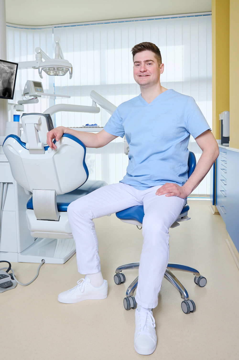
Education:
2007: Szeged University, Faculty of Dentistry
2013: Membership of ITI Danish Section (International Implantology Society)
2018: Straumann Implant Course
Ongoing: Regular annual further training courses
Speciality: General Dentistry, Dento-alveolar Surgery
Languages: Danish, English
”I chose this profession because it’s centred around people, is rooted in a desire to help, and because it’s manual, allowing me to create beautiful work. For me, it’s essential to be empathetic towards my patients. Above all, my most important professional and personal values are authenticity and quality.”
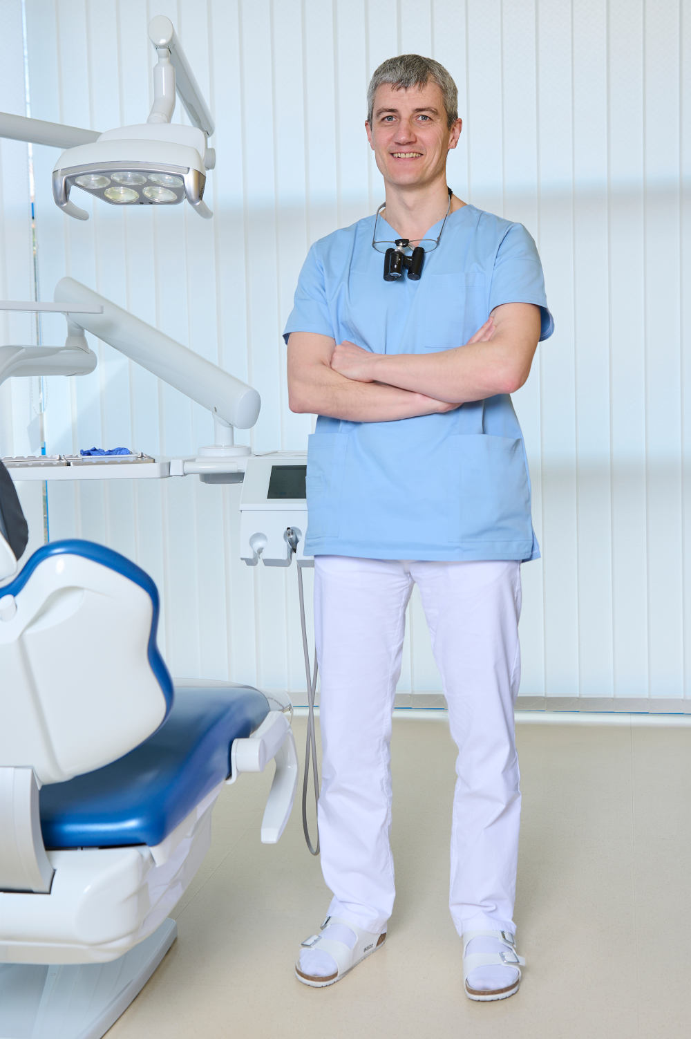
Education:
1999: Semmelweis University, Faculty of Dentistry
2001: Dental and Oral Diseases Specialisation Exam
2005: Examination in Conservative Dentistry and Prosthodontics
2018: Postgraduate Diploma of Dental Studies (Cardiff, United Kingdom)
2017: Straumann Implant Course
2018: CBCT Masterclass
Ongoing: Regular annual further training courses
Speciality: Prosthodontics, Dental Surgery (minor oral surgical procedures)
Languages: English
”From 2007 to 2019, I applied my dental expertise in Cardiff, United Kingdom. Since returning home, I’ve been utilising the knowledge I acquired at Kreativ Dental. As a proud father, I spend a significant portion of my free time with my family. Additionally, I’m heavily into sports, particularly running and swimming.”
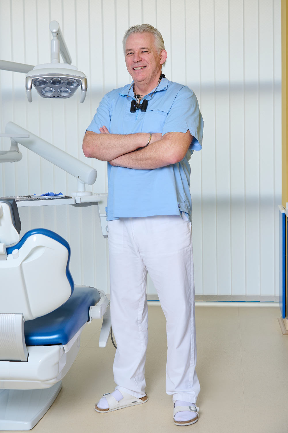
Péter examinerades 1994 som tandläkare vid universitetet i Budapest. Han fick docentstipendium i tandkirurgi under två år i Wien. Efter flera olika sjukhusanställningar med tandrestaurering och oral och maxillofacial kirurgi, som gav värdefull erfarenhet av kirurgi, kom han till vårt team 1998. Hans specialintresse är kosmetisk tandvård.
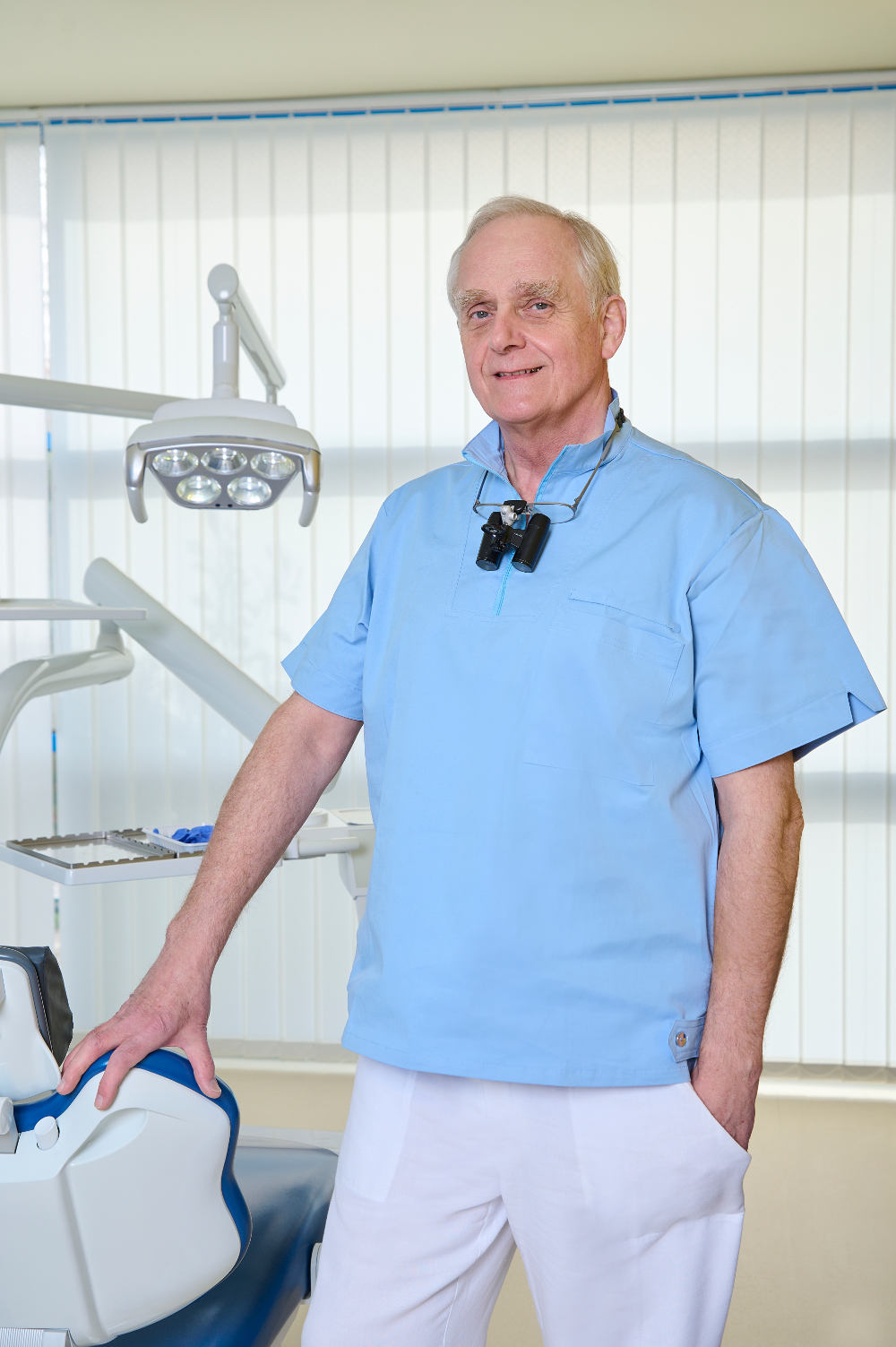
Louis examinerades först som läkare 1989 vid Semmelweis Medical University, Budapest. År 1994 examinerades han som tandläkare, och fick specialistkompetens inom oral och maxillofacial kirurgi 1999. Han har därefter arbetat enbart som tandkirurg och hans specialområde är implantatkirurgi.
Med. Reg. Nr: 48939
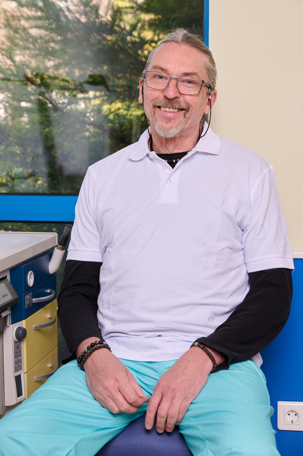
János is Leader of the Ceramic Department where all Porcelain Fused to Metal Crowns, Full Porcelain Crowns, Veneers and Inlays are skillfully crafted. Furthermore, he is one of the few exclusive demonstrators in Europe of the German company Vita which produces the best quality dental porcelain in the world.
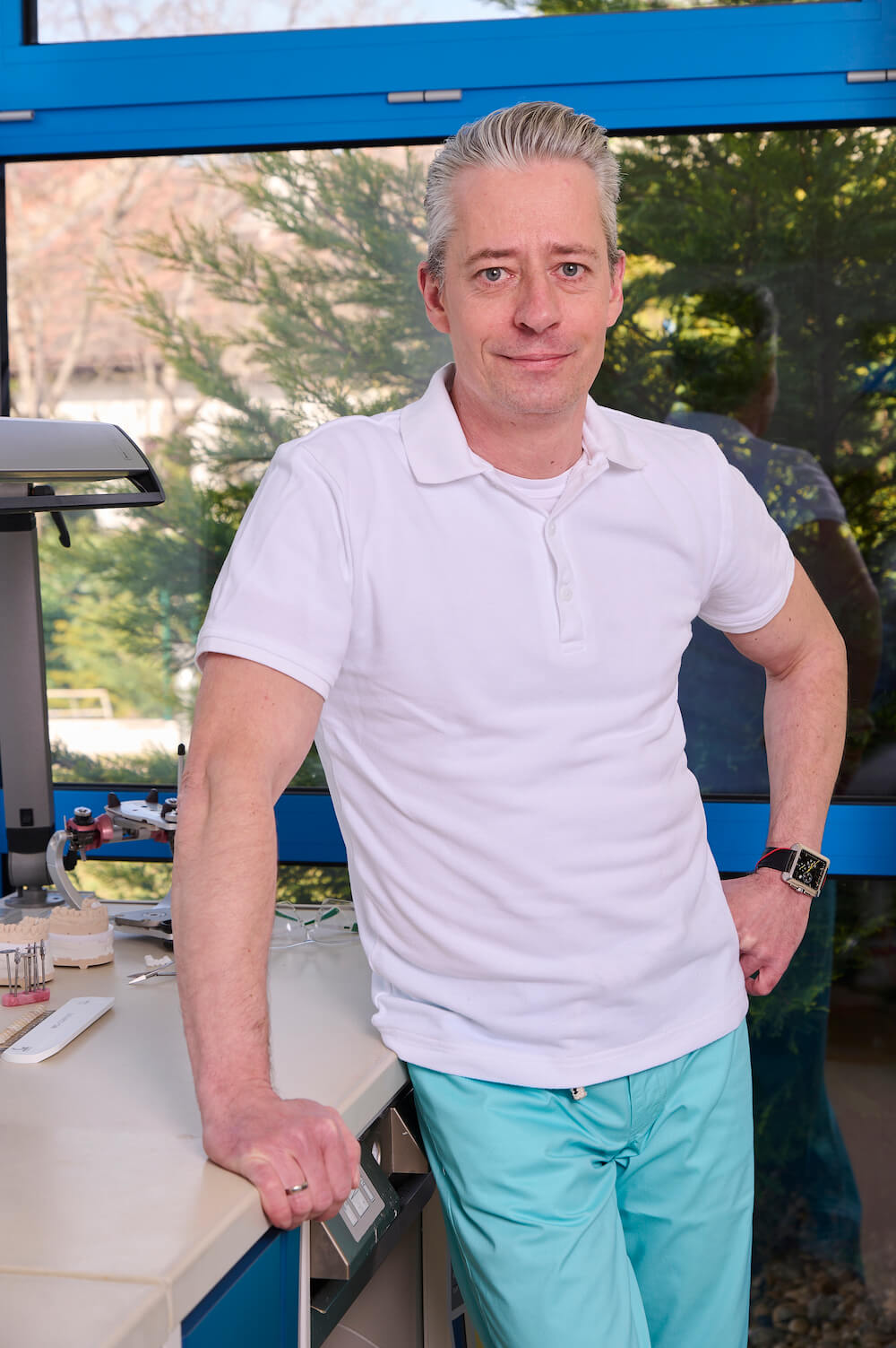
Keve Horváth joined our team of dental technicians. He works in the Ceramic Department as a master ceramist. After he graduated in 1994, he furthered his knowledge from several symposiums in Hungary, Germany and Belgium (for example Noritake and Ivoclar). In 2006 he held a course „Esthetic Front” with a real patient presence, which is a special event in Hungary and also in Europe.
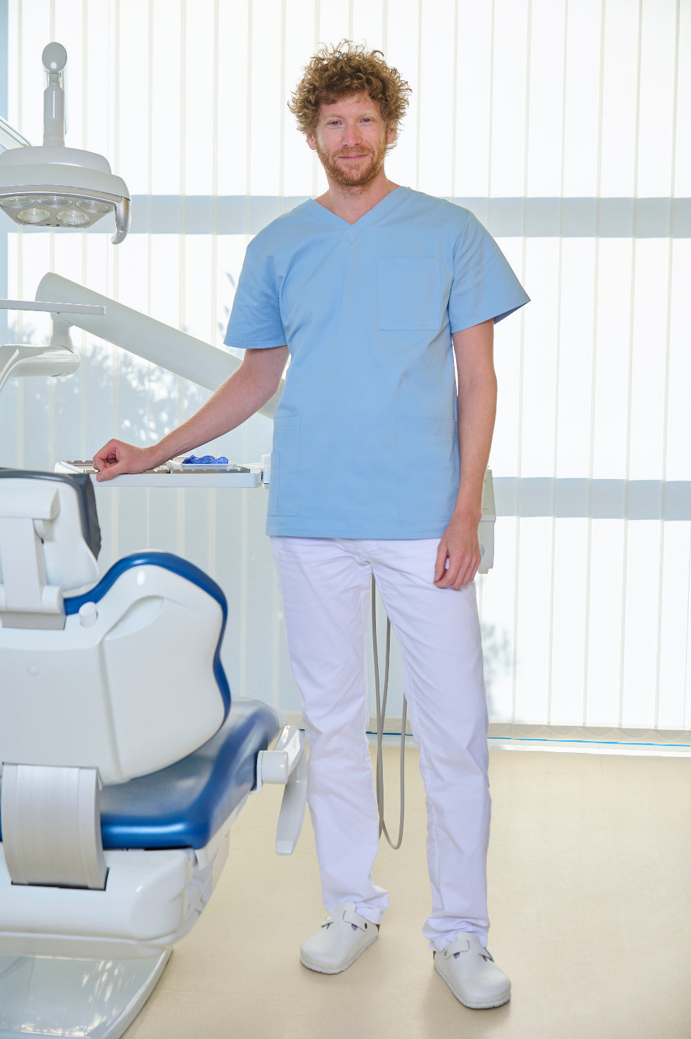
Ádám graduated in 2011 from the Semmelweis University, Faculty of Dentistry. Since then he worked at the Department of Conservative Dentistry, untill he finished a 3 year postgraduate training in conservative dentisty and prosthodontics. He also participated in the education of students both in Hungarian and English language. Ádám joined the Kreativ Dental team in 2014. His special interest is endodontics.
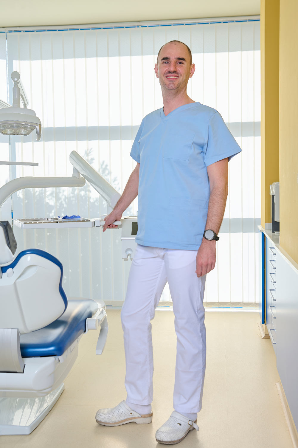
Bálint qualified at Semmelweis University, Faculty of Dentistry in 2007. Since then he has been working at the Department of Periodontology, Semmelweis University, and participates in national and international conferences (Europerio, IADR). He finished his 3 year postgraduate periodontal training in 2010. Bálint joined Kreativ Dental in 2013, his specialist field is periodontal surgery.
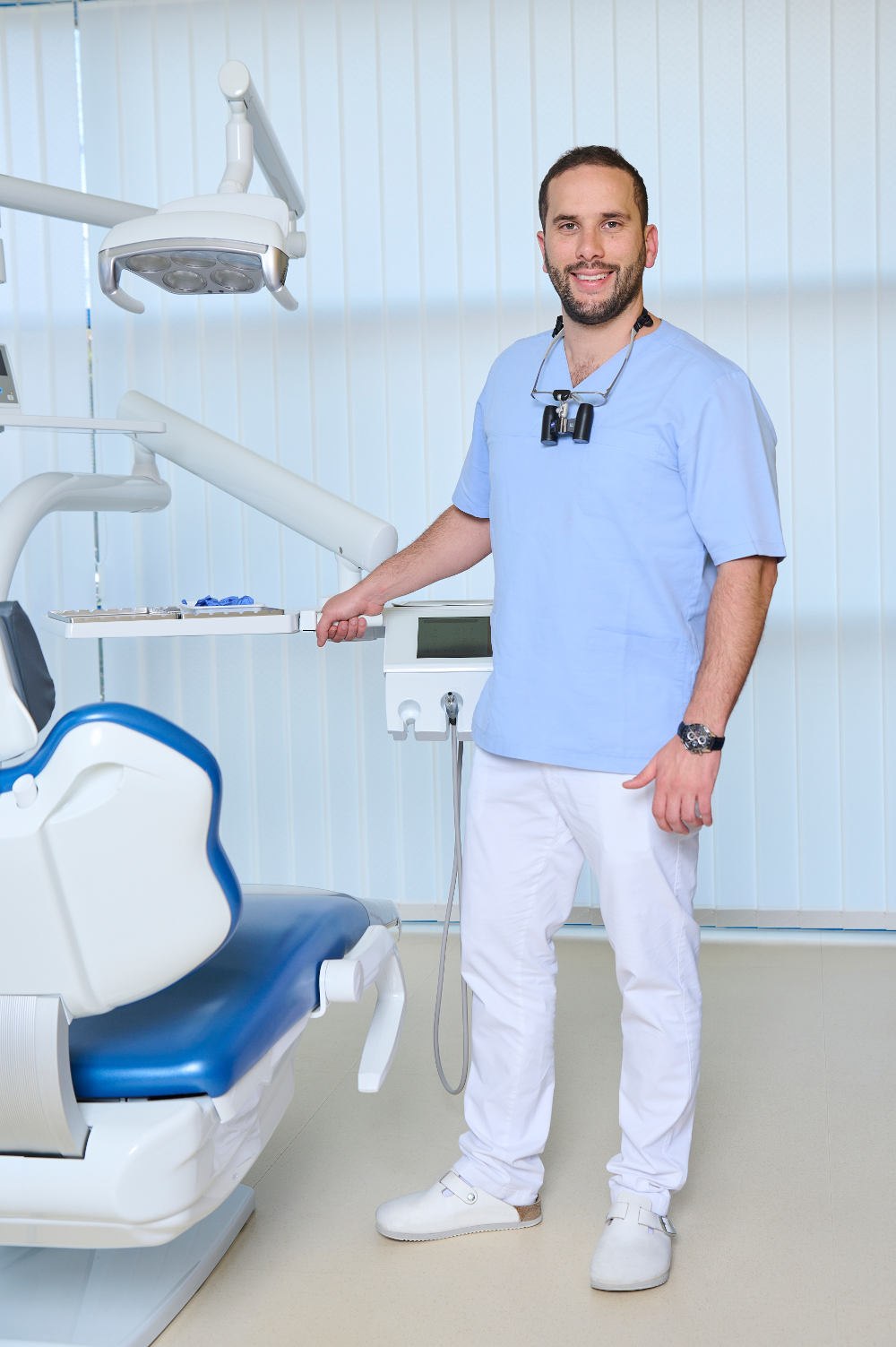
Bernard graduated in 2012 from the Semmelweis University, Faculty of Dentistry. Since then he has worked at the Department of Maxillofacial Surgery at St. John’s Hospital Budapest for 3 years and then obtained his specialization in dentoalveolar surgery in 2015. Meanwhile, he worked in private dentistry as a general dentist.
His special field is oral surgery, implantology and prosthodontics.
He joined the Kreativ Dental team in 2015.
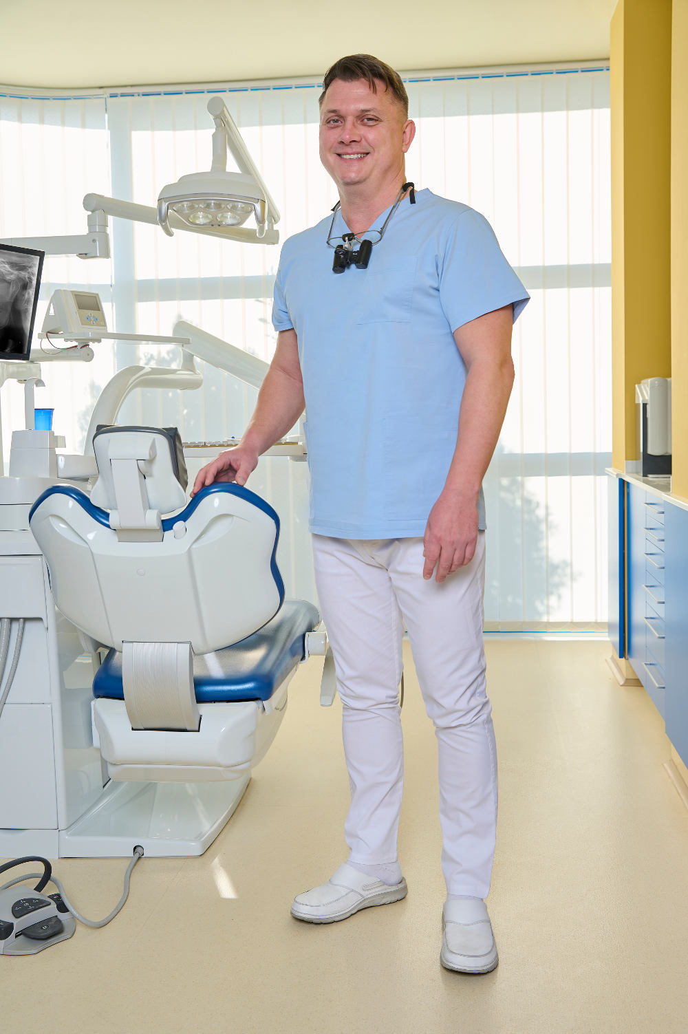
Ádám graduated in 1998 from the University of Szeged. In 2000 he became a Dental Surgeon /Specialist in General Dentistry.
After working as a Dental Surgeon at BKKMi County Hospital, Maxillo-Facial Department, Kecskemét, Hungary between 2000 and 2006, he spent 7 years as private dentist and as General Dental Practitioner in the United Kingdom.
He joined the Kreativ Dental team in 2013.
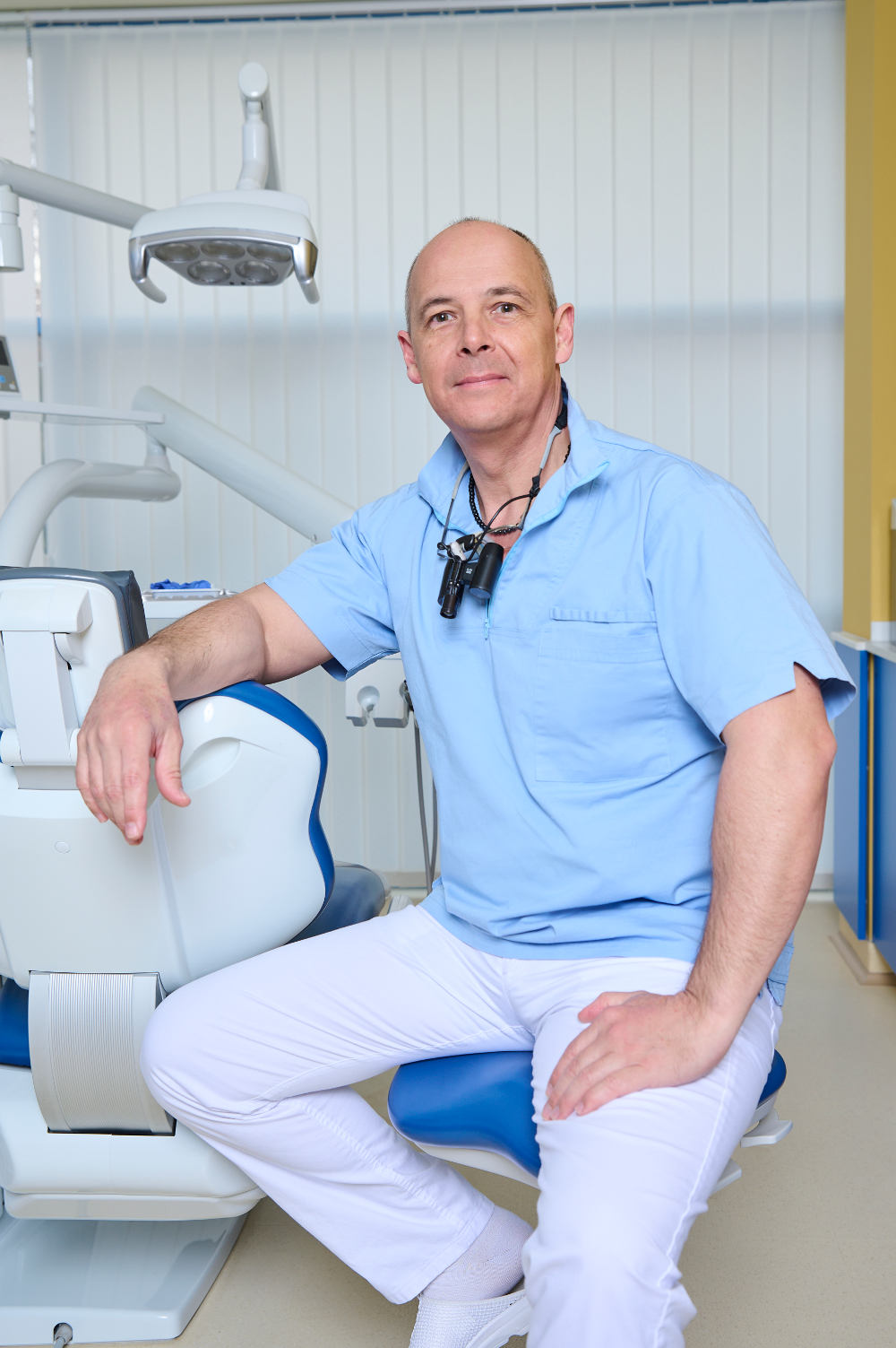
András examinerades först 1992 som tandläkare vid Semmelweis Medical University i Budapest. Efter specialistutbildning som allmäntandläkare 1995, arbetade han som privattandläkare och praktiserade som tandkirurg på Rókus sjukhuset i Budapest. Hans specialitet är kronor och bryggor.
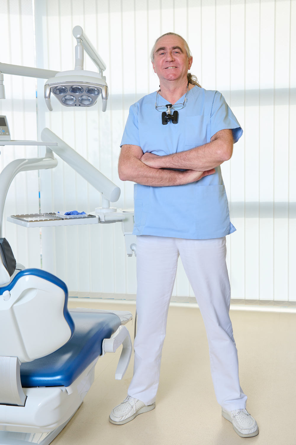
Cristian examinerades 1988 vid Neumarkt tandläkarhögskola. Han fick specialistkompetens som allmäntandläkare 1992 och har sedan 1994 gått vidare inom sina specialområden, implantat- och kosmetisk kirurgi. Cristian har också erhållit Cambridge First Certificate i engelska.
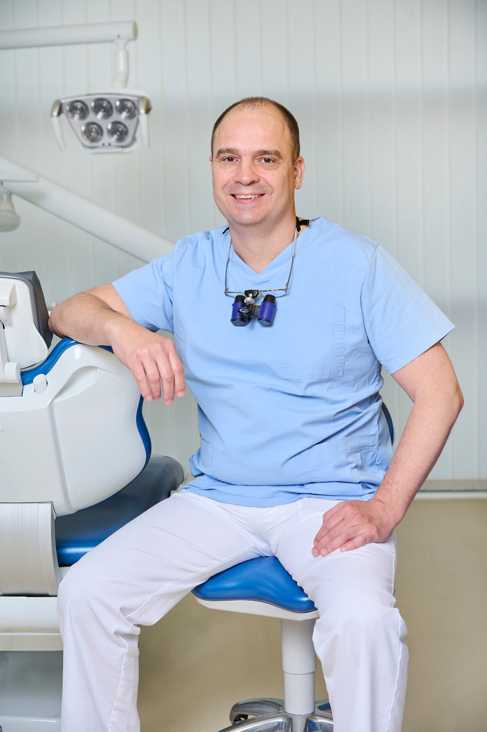
Balázs examinerades 1999 vid Semmelweis Medical University i Budapest som allmäntandläkare. Han arbetade sedan under två år som kliniker. Efter sex års erfarenhet av att arbeta med tyska och österrikiska patienter i Ungern kom han till Kreativ Dental 2007. Balázs talar både engelska och tyska.
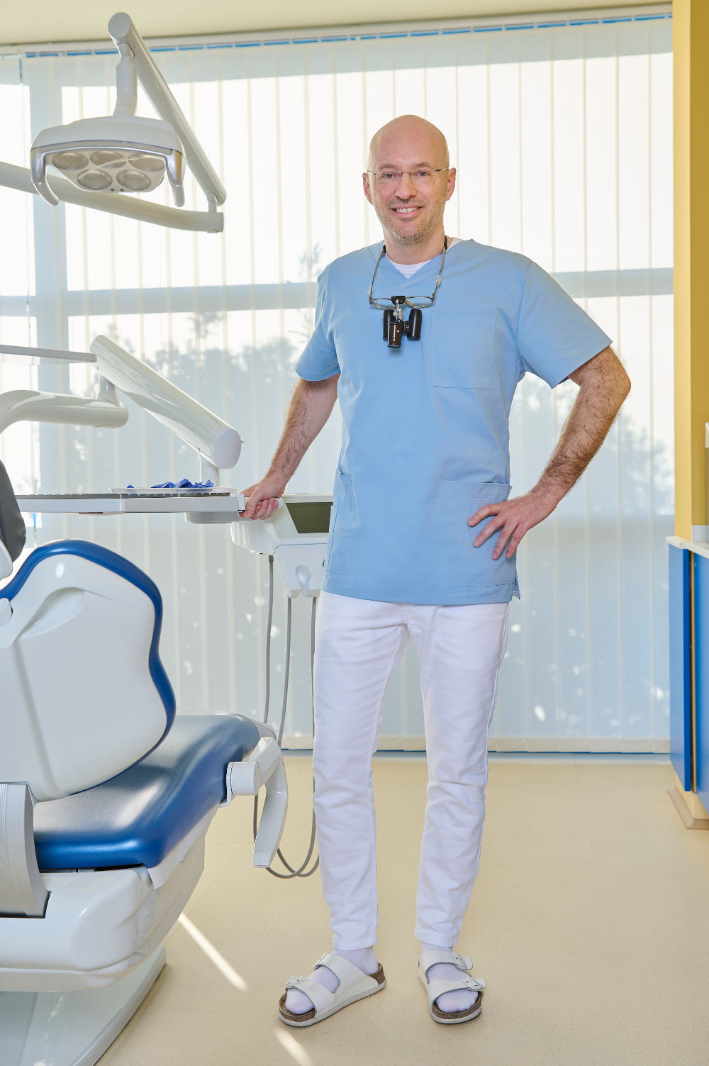
Iván examinerades 2000 vid Semmelweis Medical University i Budapest. År 2002 kom han till Kreativ Dental efter två års erfarenhet på privatklinik. Hans specialintressen är endodonti – tandrotens sjukdomar, och kosmetisk tandvård.