Dopo avere fatto ad un nostro cliente inglese una radiografia panoramica, per sostituire il dente 4, in alto a sinistra, eravamo d’accordo di inserirgli un impianto. 3 mesi fa i suoi denti sono stati estratti,per cui la mancanza è relativamente recente. L’impianto: Pitt easy V-TPS. Gli abbiamo dato un pacchetto di medicina 4 * 10 ( Dalacin antibiotici e Cataflam antidolorifici) ed abbiamo somministrato del gel-ghiaccio.
Un nostro paziente irlandese aveva perso due denti, che sono stati sostituiti con impianti 3 mesi fa. Oggi, gli abbiamo già innestato su le sovrastrutture e per la protezione delle sostituzioni dentale e abbiamo concordato con il paziente per l’applicazione di un bite ortodontico che era d’accordo con noi, quindi per il bite ortodontico abbiamo fatto anche il rilevamento dell’impronta. Siccome la preparazione di questo richiede poco tempo , sarà pronta prima della fine del soggiorno del paziente in Ungheria.
Un nostro paziente danese che da tempo aveva mancanza di denti, con l’ aiuto di impianti riceverà nuovi denti in sostituzione dei denti 4 e 6, in posizione in alto a destra. In seguito ad un sinus lifting inserendo gli impianti, gli abbiamo somministratogli antibiotici e gli antidolorifici (Cataflam Dalacin-C e TBL). Il paziente non aveva bisogno di una protesi provvisoria. La prossima volta – prevedibilmente fra 4 mesi – il paziente può ottenere nuovi denti.
63 enne paziente di sesso femminileha chiesto gli impianti al posto dei denti in alto a destra quelli 3, 5, e 6 che erano mancanti da qualche tempo. All’ ‘anziana signora sembrava chiaramente di averespanso la cavità del seno, per cui lo strato osseo che serviva per la presa dell’ impianto era troppo sottile. Prima dell’impianto, eseguiamo il sollevamento del seno affinché l’osso dia una struttura fissa per gli impianti.
An American female patient presented with multiple missing teeth and an unremarkable resorbtion in the jaw ridge. In order to be able to place implants in positions 23,24,25,26 it was necessary to carry out a minimal sinus lift. The sinus lift is a surgical treatment which we usually carry out with local anaesthetic. In the case of this patient we used BioOss, a bone replacement material of animal origin. Because the sinus lift was not significant we carried out the bone replacement and placing of implants in only one session. We also used BioGide membrane to help the tissue to heal quicker. The collagen membrane, also of animal origin, helps the wound to heal and has a positive effect on bone regeneration as well. We will remove the patient’s stitches next week. Then a 6 month osseointegration period is necessary before we will finally be able to place the new porcelain teeth. Unusually, the lady did not want a temporary denture/crowns for the interim period.
A returning middle-aged Hungarian gentleman complained of sensitive gums which bled easily when pressure was applied in the area of tooth 47 where a dental implant was inserted many years ago. During the examination we diagnosed inflammation and periimplantitis around the implant.
The inflammation around the implant had penetrated to deeper levels and had caused the bone tissue to disappear. The cause of an infection of this nature is usually bacterial. In the pockets which had developed between the dental implant and mucosa, with a lack of regular and thorough mouth and tooth care the bacteria can easily colonize and cleaning off the tartar can be difficult. If the infection also reaches the bone tissue the implants can loosen and if left untreated the patient may lose them. Our patient’s inflammation around the implant was indeed at an advanced stage. The tissue decay had loosened the implant as well therefore we removed it. After proper cleansing of the wound we carried out a new implantation. The patient will get a new crown after the healing period.
Un nostro paziente 26 enne ci ha visitato a causa di due denti incisivi superiori rotti in un incidente d’infanzia, che sono stati costruiti con otturazioni in composito. Visto che questa soluzione non era per lui esteticamente appropriata, preferiva avere le corone. Abbiamo fatto una radiografia panoramica, consegnandogli un piano di gestione. Il piano è stato accettato da lui. I due denti incisivi, con lucidatura sono stati preparati da noi per le corone in zirconio, inoltre abbiamo fatto anche il rilevamento diprecisione dell’impronta.
Ad un giovane paziente norvegese abbiamo applicato corone di zirconio nell’arcata superiore (da 3 a 3 ). Dato che per lui era molto importante l’aspetto estetico, per la parte frontale ha scelto lo zirconio, più costoso ma più gradevole, mentre gli altri denti (quelli superiori e inferiori da 4 a 7) sono stati ricostruiti con corone in metallo-ceramica. La ricostruzione è stata fatta con successo, sia funzionalmente che esteticamente. Il paziente era contento.
Un nostro paziente, un uomo di mezza età, è tornato alla clinica a causa di un precedente danno alla corona del suo ponte in ceramica. Dato che il problema si è verificato di notte, per proteggere il ponte circolare superiore e inferiore, gli abbiamo proposto l’uso di un bite durante la notte. Non trattandosi di un digrignamento grave, abbiamo ritenuto sufficiente l’utilizzo di una guida piuttosto sottile, costruita dopo aver eseguito un rilevamento dell’impronta. Per prepararla ci sono voluti 1-2 giorni.
52 enne signora norvegese è arrivata per farsi la prova di struttura. Le sarà preparata una sostituzione di denti, da quelli in alto a destra 7 a quelli di 7 in alto a sinistra. Consultandoci con la paziente, abbiamo determinato anche il colore del dente. Siccome la mancanza dei denti si è estesa sull’intera dentiera superiore, la paziente aveva la possibilità di scegliere un colore leggermente diverso da quello originale. Visto che la signora aveva dei denti inferiori incompleti, le abbiamo posto due ponti laterali nella parte inferiore.
Un uomo di media età é venuto nel nostro studio con un dente mancante nell’arcata della dentatura di sinistra. Il posto del dente mancante era ancora fresco, appena atrofizzato, ciò é caratteristico di un tratto di gengiva privo di stress per lungo tempo. In occasione del primo trattamento abbiamo eseguito la levigatura, il rilevamento dell’impronta dentale ed applicato una corona provvisoria. A tal punto abbiamo fatto una prova per la struttura che serviva al ponte in metallo ceramica, rispettivamente abbiamo fatto anche un nuovo rilevamento dell’impronta dentale tramite Futar-D. La prova è riuscita, l’altezza è perfetta.
One of our American patients presented a few days previously with dental restorations in a bad condition. His/her expectationwas that we would produce an upper and lower round bridge on his/her existing implants. At the first treatment we prepared the teeth in the lower jaw, grinded them down, exposed the implant sites and took a precision impression. Today we prepared the left 3 in the upper jaw. We exposed the implants existing int hat region and placed impression caps into them. We then took a precision impression. Following the production of the round bridges at the next treatment the round bridges were fitted to the upper and lower implants. After permanently fitting the bridges, the patient could smile again!
A 23 year old Irish patient presented following many previous dental treatments. We found 3 well placed implants in the patient’s upper jaw, but the abutments and many of the young lady’s own teeth were in an eroded and abraded condition. We fitted zirconium and porcelain fused to gold crowns and bridges, and glass fibre post cores, to the implants and the patient’s own teeth. Here we were able to save 2 roots which were still in a good condition. The patient’s teeth show that during previous treatment acute complaints were indeed carefully attended to but no attempt was made to solve the probable causes of the problems: nocturnal grinding (bruxism). On our recommendation the young lady also received a nightguard (bite raising splint for night time use).
An upper porcelain fused to metal round bridge with post cores was produced swiftly within 1 week in the dental technical laboratory for a 60 year old English female patient. For the fitting and cementations we used FujiPlus cement. The height of the bite was fine. We took an alginate impression for the patients bite raising nightguard. The patient can collect the nightguard in 2 days’ time. We provide the patient with further oral hygiene advice. She needed this on the basis of the condition of the oral cavity. The patient was very satisfied with the treatment and had the opportunity to see the sights of Budapest whilst waiting for her new teeth.
A 32 year old Norwegian lady came to us following her second pregnancy as her dentition had deteriorated significantly following the births. We prepared all of her upper teeth, took impressions of the tooth stumps and produced a temporary bridge.
The patient wanted full porcelain crowns, where the crown frame is also made from zirconium-oxide. In this instance we produce the frame using 3D computer scanning techniques which enables better precision and aesthetic results in the finished product. The laboratory procedure is therefore more time consuming and the materialas well as the method make the process more expensive but the look of the crowns is very important to this Norwegian patient.
Ad un 40 enne paziente francese abbiamo praticato il trattamento del canale radicolare sul dente 2 in alto a sinistra. La lunghezza del canale radicolare è stata determinata da noi tramite strumenti di misurazione. Il canale radicolare richiedeva una dilatazione meccanica. Dopo aver eseguito la disinfezione e asciugatura del canale radicolare, l’otturazionedel canale radicolare l’abbiamo preparata con del Sealapex , mentre l’otturazione di copertura è stata realizzata con cemento Fuji IX.
One of our Norwegian male patients complainedof a recurring sometimes splitting toothache. During the diagnosis we established that tooth 32 was significantly decayed and a root canal treatment would be necessary. During the treatment the root canal of tooth 32 was opened up and cleaned using WDW Gold. During the rinsing of the root canal we used Solumium then placed a medical (calcipulp) temporary filling. As expected the patient’s pains subsided and disappeared gradually after a few days. If everything goes well we will change the temporary root filling for a permanent one in 8 days’ time.
Un nostro paziente ungherese è arrivato con infiammazioni gengivali. Due mesi fa, ha ricevuto un impianto al posto del dente 4 in alto a destra. L’infiammazione era tra i denti 4 e 3 in alto a destra. Le radiografie degli impianti sono state controllate e trovati bene. Al fine di disinfezione dell’infiammazione, la parte della gengiva infiammata è stata lavatacon Betadin; le gengive del paziente molto probabilmentesono state ferite da un cibo duro, ma nonostante ciò, non abbiamo trovato altri cambiamenti patologici.
Un nostro paziente ungherese è stato operato in Germania, dove gli hanno estratto il dente 8 in basso a sinistra (dente di giudizio), anche perché – secondo il rapporto del paziente – visto che questo era circondato da un ascesso. È arrivato con il viso gonfiato lamentando di un dolore lancinante. Abbiamo fatto fare sciacqui con Betadinos Hyperolos quindi e rimosso i frammenti di osso dalla ferita. Infine, gli abbiamo somministrato degli antibiotici. Se tutti i frammenti di osso sono stati rimossi correttamente, il dolore se ne andrà in un periodo di tempo molto breve ed anche il gonfiore diminuirà presto.
Un 31 ennepaziente è arrivato con un mal di denti crescente da giorni. Inoltre, lui ha scoperto una decolorazione visibile sulla dentatura inferiori e sui molari anteriori. La diagnosi del paziente era corretta, davvero si era di fronte a denti forati. Per il dente 4 in basso a destra e quello asinistra abbiamo preparato un’ otturazione una foto- polimerizzazione , che ha il vantaggio diindurirsi immediatamente al contatto con la luce, senza necessità di dovere aspettare neanche 1-2 minuti, ed inoltre possiede eccellenti proprietà estetiche.
Un giovane uomo paziente norvegese ha dormito male per molto tempo, si è svegliato stanco, con il mal di testa, e il collo rigido Inoltre, aveva regolarmente le gengive infiammate, dolori nei muscoli masseteri, un segnale, che ha dormito l’intera notte a denti stretti. Ciò è stato anche confermato dalle tracce di usura, che abbiamo scoperto su alcuni denti . All’uomo abbiamo consegnato un bite ortodontico da noi preparato che gli ha impedito il permanente morso dei denti proteggendoli dall’usura.
29- enne paziente da anni si lamenta per mal di denti, non insopportabile, ma fastidioso. Nel suo dente, in alto a destra abbiamo trovato una profonda carie dove in occasione di un’applicazione indiretta del cappello di polpa abbiamo utilizzato il disinfettante Dycelal coprendone poi di cemento-vetro ionomero Fuji II.LC. Poiché l’otturazione era provvisoria, il paziente doveva tornare dopo 3 settimane per vedere se gli agenti patogeni fossero estinti o se fosse necessario il trattamento di radice qualora fosse avvenuta l’infiammazione della polpa.
The ortho-pantograph x-ray of our 50 year old Italian femaile patient showed that her upper right 6 and upper left 6 (or rather their remains) needed extracting and replacing. The extracted teeth were replaced with bridgework according to the plan. In the first stage of treatment we prepared the right and left upper 5 and 7 teeth, and removed the right and left upper roots (radixes). We took impressions for the dental laboratory and produced temporary crowns. The dental technical laboratory will produde the final crowns in under 1 week and the Italian lady’s next visit will be due in only 2 weeks’ time. This way she was able to purchase an economy flight ticket.
A young Swiss man complained of a sharp throbbing pain in his lower left teeth. The problem was caused by the severely inflamed 37. We trepanated and extirpated this number 7, which means we opened the pulp chamber and removed the inflamed nerves. We carried out the widening and cleaning with WDW Gold. We placed a Sealapex root filling and then a Fuji IX tooth filling.
A 60 year old English gentleman presented with many problems. Firstly we surgically removed teeth 12,44,45 and 48. We produced the treatment plan for the replacement of the teeth but he did not want to begin the restoration treatment on this visit.
Secondly in the right upper and lower quadrants it was necessary to remove tartar. Following the ultrasound treatment and cleaning we carried out a curettage in the deeper parts of the gum pockets. This is necessary because the deeper parts of the gum pockets are not reachable with ultrasound scaling. With the help of the appropriate instruments these areas can be cleaned for which local anaesthetic is usually required. On finishing treatment we provided our patient with oral hygiene advice for which the patient was especially grateful.
Kreativ Dental Clinic © 2025
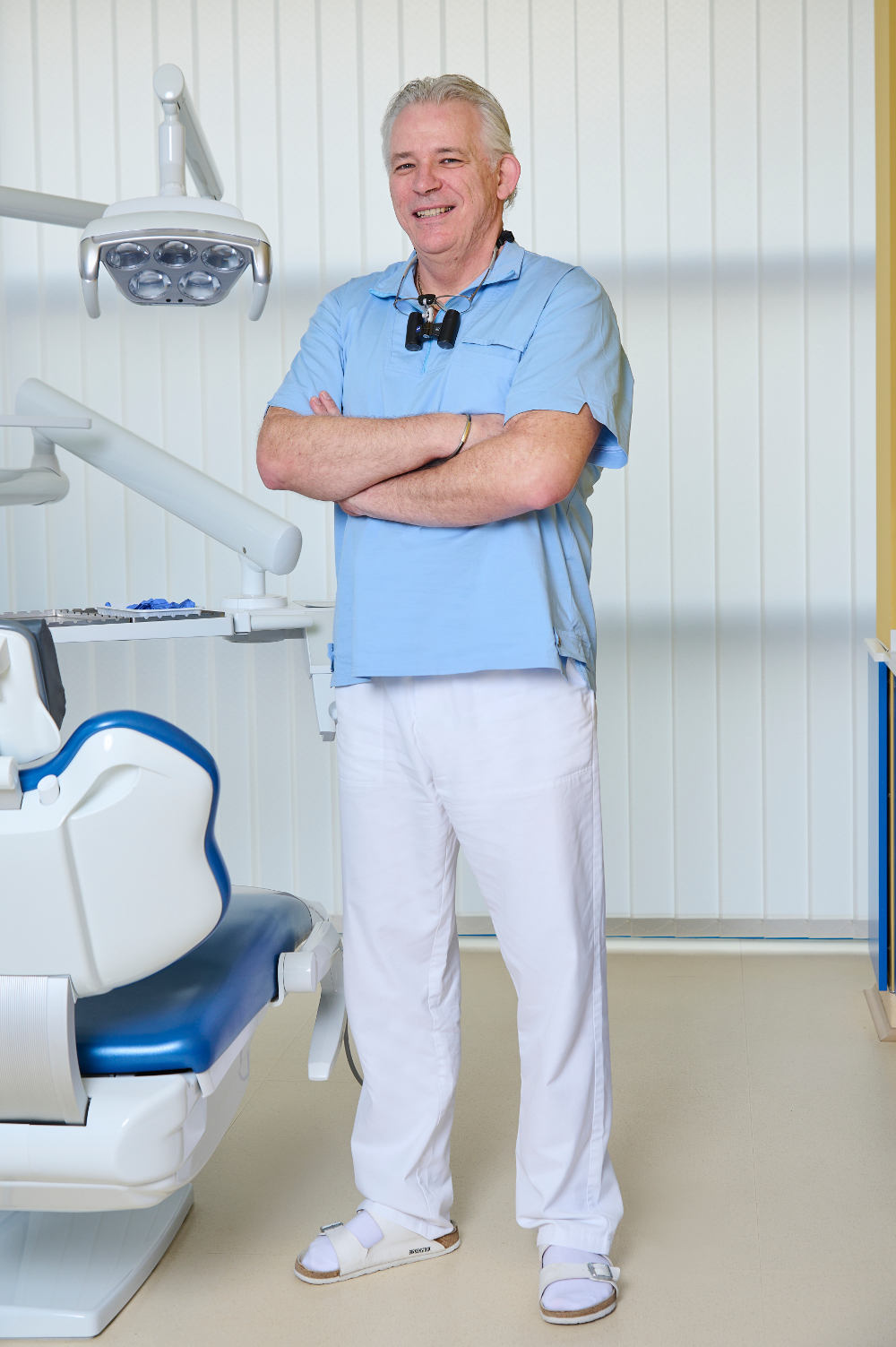
Péter si è laureato nel 1994 presso l’Università di odontoiatria di Budapest. Ha ottenuto una borsa di studio in Chirurgia dentale di due anni nell’ Univeristà di Odontoiatria di Vienna. Dopo diverse posizioni ospedaliere nell’Odontoiatria Ristorativa e Chirurgia orale-seno paranasale, dove ha acquisito un’importante pratica chirurgica, nel 1998 ha aderito al nostro team Odontoiatrico. La sua specializzazione è l’odontoiatria estetica.
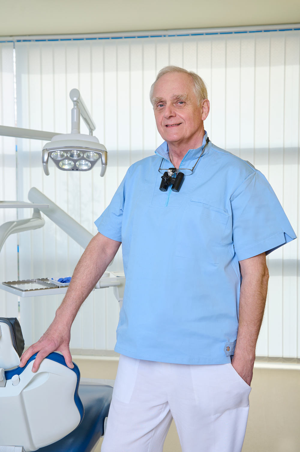
Lajos, prima si è laureato nel 1989 come Medico generale presso l’Università di Medicina Semmelweis di Budapest. Poi nel 1994 si è laureato come Odontoiatra generale, ed ha ottenuto la specializzazione in Chirurgia Maxillo-facciale nel 1999. Dal 1999, lavora esclusivamente come Chirurgo dentale. Il suo campo di specializzazione è l’implantologia odontoiatrica.
Medical Registration Number: 48939
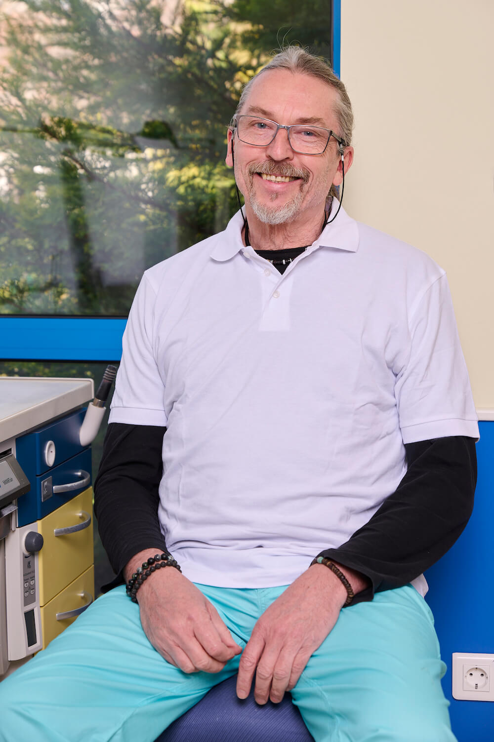
János è il Leader del Dipartimento di odontotecnica, dove sono prodotte tutte le Corone in Porcellana fusa in metallo, le Corone in Porcellana integrale, le faccette e gli Inlay. Inoltre, è uno dei pochi rappresentanti esclusivi in Europa dell’azienda tedesca Vita, che produce la Porcellana odontoiatrica dalla migliore qualità nel mondo.
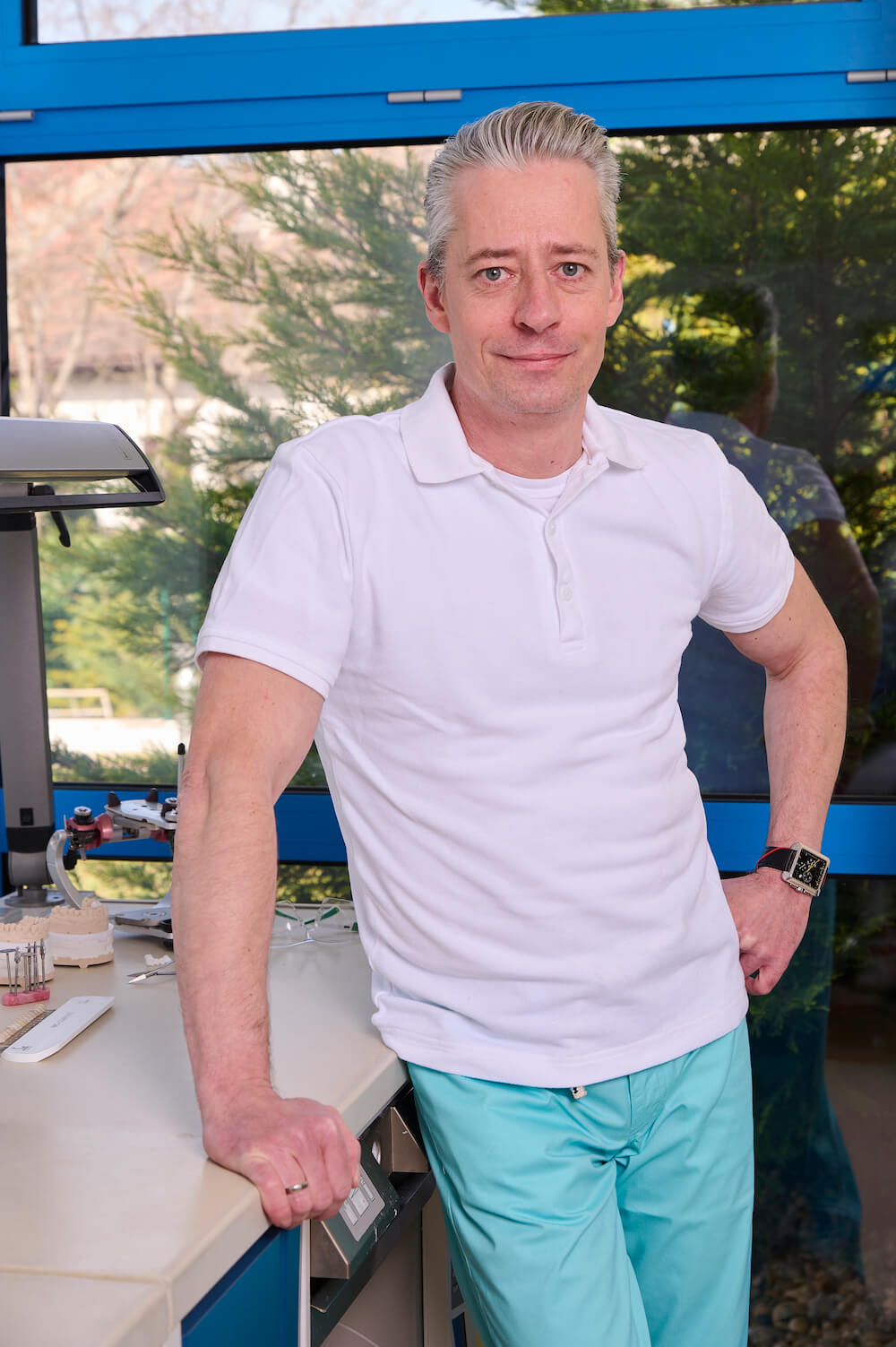
Keve Horváth joined our team of dental technicians. He works in the Ceramic Department as a master ceramist. After he graduated in 1994, he furthered his knowledge from several symposiums in Hungary, Germany and Belgium (for example Noritake and Ivoclar). In 2006 he held a course „Esthetic Front” with a real patient presence, which is a special event in Hungary and also in Europe.
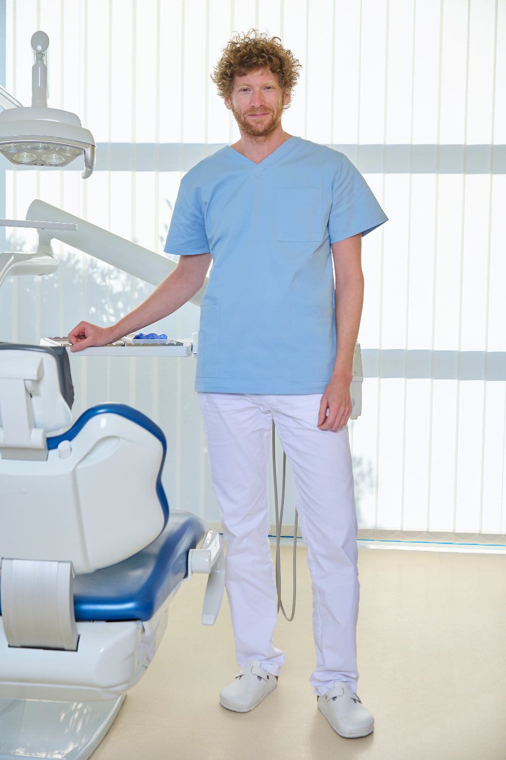
Ádám graduated in 2011 from the Semmelweis University, Faculty of Dentistry. Since then he worked at the Department of Conservative Dentistry, untill he finished a 3 year postgraduate training in conservative dentisty and prosthodontics. He also participated in the education of students both in Hungarian and English language. Ádám joined the Kreativ Dental team in 2014. His special interest is endodontics.
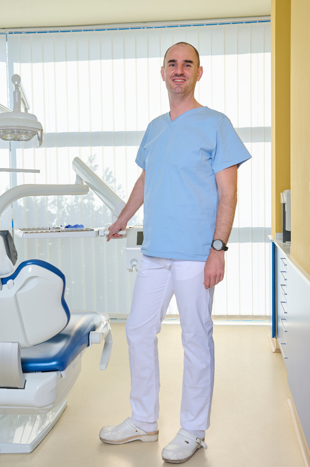
Bálint qualified at Semmelweis University, Faculty of Dentistry in 2007. Since then he has been working at the Department of Periodontology, Semmelweis University, and participates in national and international conferences (Europerio, IADR). He finished his 3 year postgraduate periodontal training in 2010. Bálint joined Kreativ Dental in 2013, his specialist field is periodontal surgery.
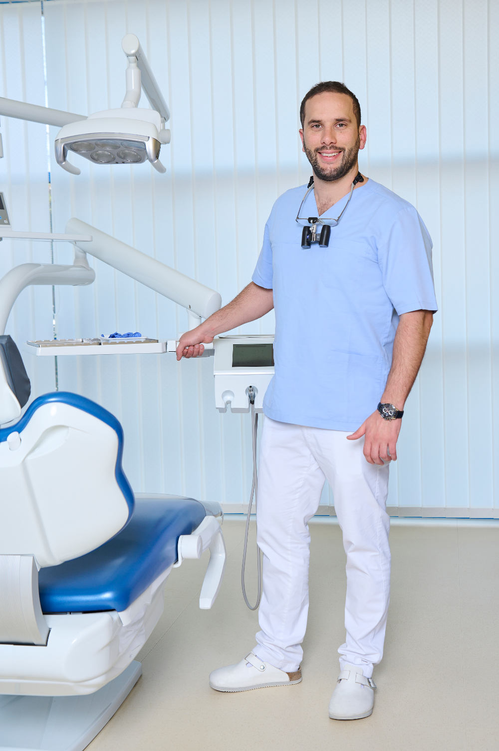
Bernard graduated in 2012 from the Semmelweis University, Faculty of Dentistry. Since then he has worked at the Department of Maxillofacial Surgery at St. John’s Hospital Budapest for 3 years and then obtained his specialization in dentoalveolar surgery in 2015. Meanwhile, he worked in private dentistry as a general dentist.
His special field is oral surgery, implantology and prosthodontics.
He joined the Kreativ Dental team in 2015.
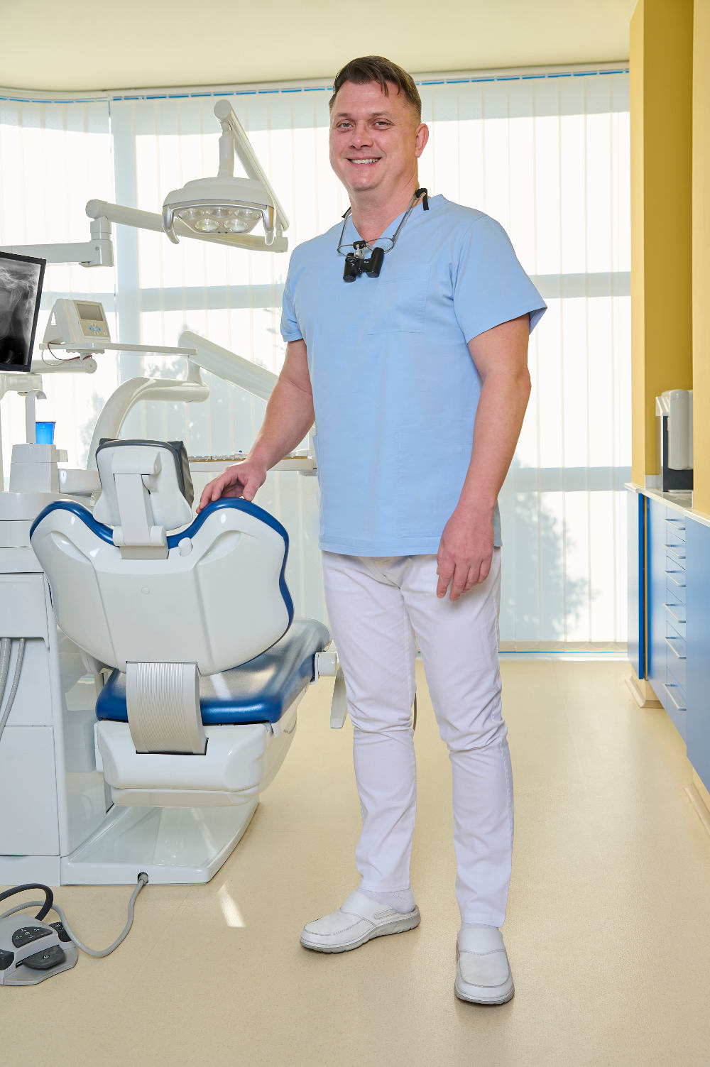
Ádám si è laureato nel 1998 presso l’Università di Szeged. Nel 2000 divenne Dentista / Specialista in Odontoiatria Generale.
Dopo aver lavorato come Denista presso BKKMi County Hospital, Dipartimento di Chirurgia Maxillo-Facciale, di Kecskemét, in Ungheria tra il 2000 e il 2006, ha trascorso sette anni come dentista privato e come dentista praticante nel Regno Unito.
Si è unito al team Kreativ Dental nel 2013.
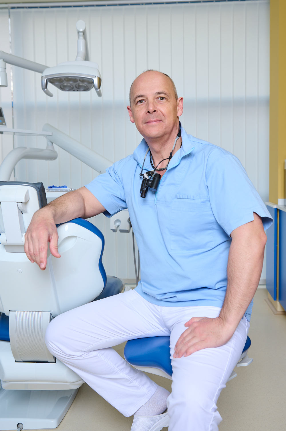
András si è laureato nel 1992 come Odontoiatra presso l’Università Semmelweis di Budapest. Da allora, ha lavorato presso Cliniche Private e come praticante presso la Chirurgia Odontoiatrica dell’ospedale Rókus di Budapest. Le sue specializzazioni sono le Corone e i Ponti.
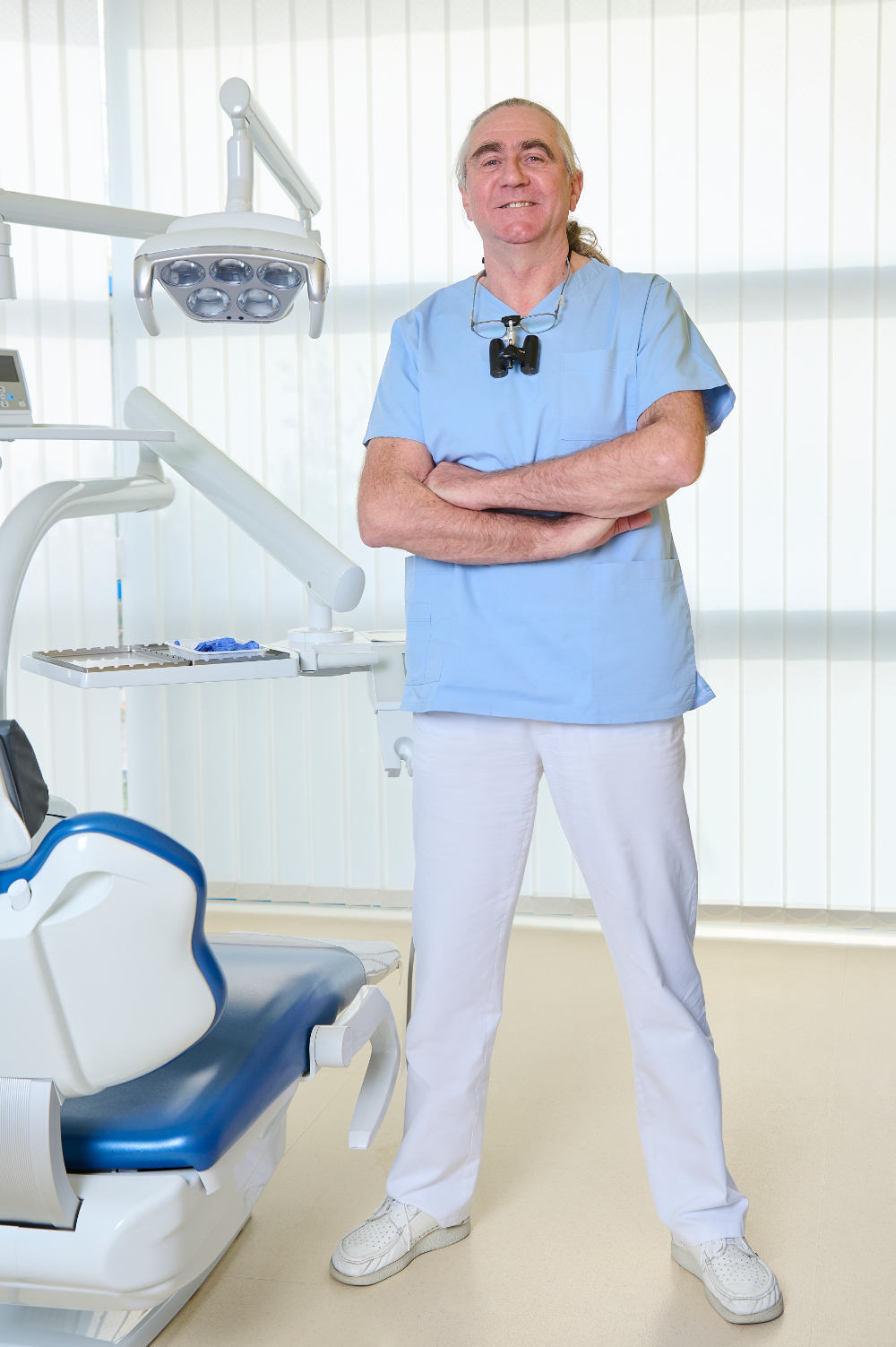
Cristian si è diplomato nel 1988 presso la Scuola Odontoiatrica Neumarkt. Nel 1992 si è laureato in Odontoiatria. Dal 1994 si è dedicato all’Implantologia e all’Estetica Dentale. Cristian ha ottenuto il certificato Cambridge First Certificate di inglese.
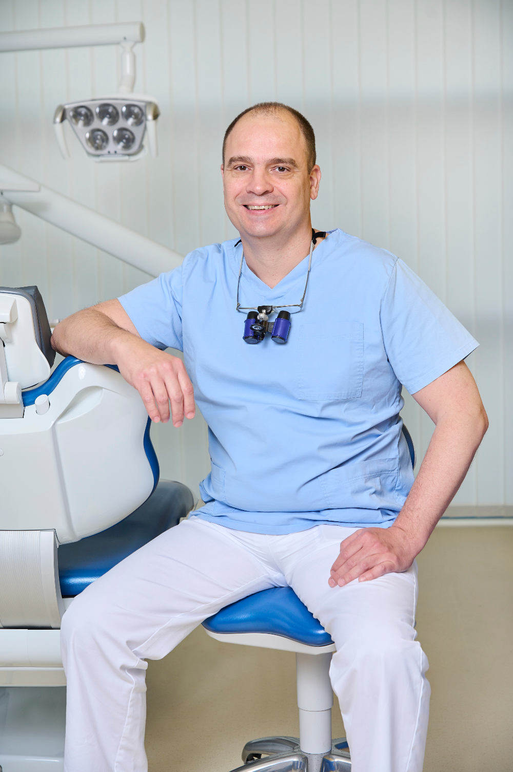
Nel 1999 Balázs si è laureato in Odontoiatria presso l’Università di Medicina Semmelweis di Budapest. Nel 2007, dopo 6 anni trascorsi operando pazienti tedeschi e austriaci presso altre cliniche dentistiche private, è entrato a far parte del team Kreativ Dental.
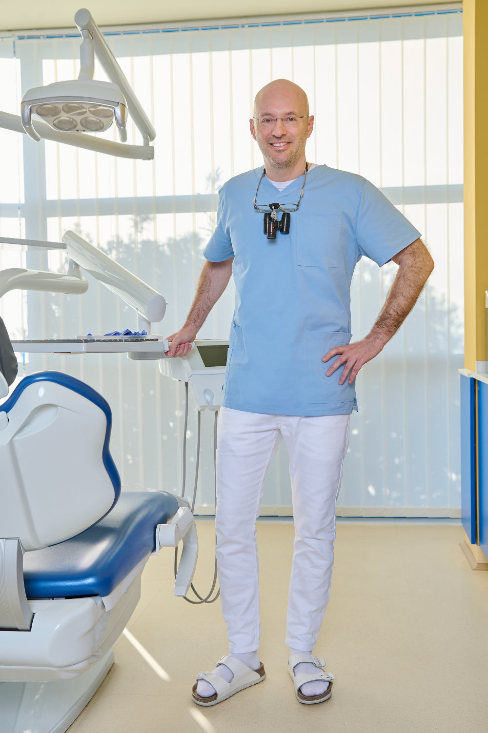
Iván si è laureato in Odontoiatria nel 2000 presso l’Università di Medicina Semmelweis di Budapest. Dopo due anni trascorsi in altri Studi Privati, nel 2002 è entrato a far parte dello staff Kreativ Dental. Le sue specializzazioni: Endodonzia, la cura delle patologie della polpa dentaria e l’Estetica Dentale.